A tumor classification system based on radiomics

A radiomics and tumor technology, applied in the field of medical image processing, can solve the problems of physical trauma, great difference in degree of differentiation, incomplete consistency of molecular typing and immunohistochemical detection results, etc., to achieve comprehensive and reliable information considerations good repeatability

Active Publication Date: 2021-01-12
NORTHEASTERN UNIV LIAONING
View PDF5 Cites 2 Cited by
  • Summary
  • Abstract
  • Description
  • Claims
  • Application Information

AI Technical Summary

Problems solved by technology

Clinicopathological diagnosis generally uses puncture or surgical biopsy, which not only causes trauma to the body, but also shows certain limitations. The pathological morphology of the same region may be different, and the degree of differentiation may vary greatly in different regions; similarly, the heterogeneity of breast cancer It may also make the molecular typing and immunohistochemical detection results obtained by gene chip technology not completely consistent

Method used


Image

  • A tumor classification system based on radiomics
  • A tumor classification system based on radiomics
  • A tumor classification system based on radiomics

Examples


Embodiment Construction

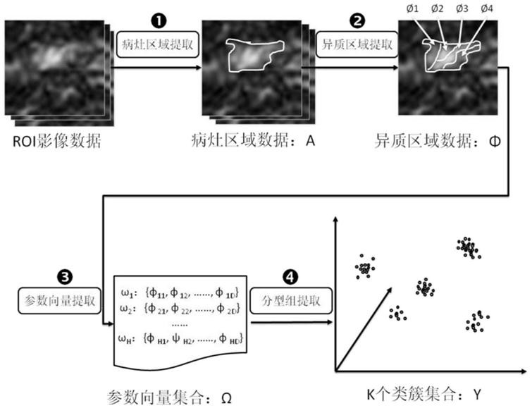
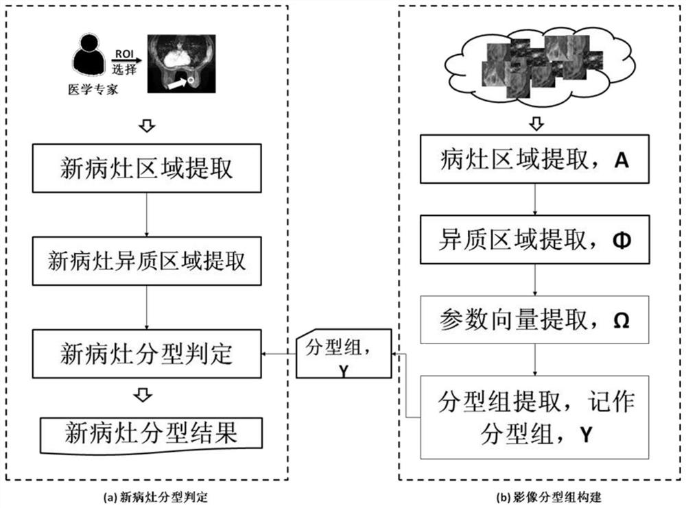
[0050] The construction process of the image classification model of the present invention is as follows: figure 1shown. This process is divided into two processes: the process of image typing group construction and the process of new lesion typing, in which the operations of extracting lesion areas and extracting heterogeneous areas are the same in the above two processes.

[0051] 1. The process of image typing group construction

[0052] The image typing group construction process is as follows: figure 1 (b) shown. Based on a large number of breast cancer ROI (Region of Interest) image data, first extract the lesion area data A, and then extract heterogeneous areas for data A, extract texture, dynamics, shape, statistics and other parameters for each heterogeneous area to construct multi-dimensional The similarity clustering method is used to divide these heterogeneous regions into K sets, and each cluster set is recorded as an image typing group, and the center of K set...


PUM


Abstract

The present invention provides a radiomics-based tumor classification system, including: lesion area extraction, used to extract lesion data; heterogeneous area extraction, using a clustering method to extract all initial heterogeneous areas in the lesion data, and then Determine the final clustering result based on the connectivity and number of pixels of the clusters on the image, and find out multiple heterogeneous regions; parameter vector extraction, extract the shape, state and other parameters of each heterogeneous region; type group extraction, The clustering method is used to divide each heterogeneous area into clusters to obtain multiple categories. New lesion type determination is used to determine which type each heterogeneous type of new lesion belongs to. The new lesion typing result is used to output the new lesion typing result. The tumor typing system of the present invention has the advantages of non-invasiveness, non-intervention, repeatability, etc., and its analysis object is based on all lesion tissues, and the information is comprehensive.

Description

technical field [0001] The invention belongs to the technical field of medical image processing, and in particular relates to a radiomics-based tumor classification system. Background technique [0002] With the prolongation of the average life expectancy of human beings, the threat of cancer to human beings has become increasingly prominent, and it has become the first cause of death of urban and rural residents in our country. Taking breast cancer as an example, breast cancer refers to malignant tumors occurring in breast ductal epithelium. It is one of the most common malignant tumors in women, with an incidence rate of 23 / 100,000, accounting for 7-10% of all kinds of malignant tumors in the whole body. Between the ages of 45 and 55, and showing an increasing trend year by year, especially Shanghai, Beijing, Tianjin and the coastal areas are the high incidence areas of breast cancer in my country. The establishment of breast cancer screening and early diagnosis system can...

Claims


Application Information

Patent Timeline
Patent Type & Authority Patents(China)
IPC IPC(8): G06T7/00G06K9/62G06T7/11G06T7/194G06T7/66G06T7/187
CPCG06T7/0012G06T7/11G06T7/187G06T7/194G06T7/66G06T2207/30096G06F18/23
Inventor 栗伟于鲲冯朝路郭志伟王东杰赵大哲
Owner NORTHEASTERN UNIV LIAONING
Why Eureka
Try Eureka