Medical image-processing apparatus, x-ray ct apparatus, and medical image processing method

A medical image and processing device technology, applied in image data processing, image enhancement, image analysis, etc., can solve problems such as insufficient diagnosis and difficulty in confirming lung function reduction

Pending Publication Date: 2018-11-09
CANON MEDICAL SYST COPRPORATION
View PDF12 Cites 1 Cited by
  • Summary
  • Abstract
  • Description
  • Claims
  • Application Information

AI Technical Summary

Problems solved by technology

However, for early detection of lung diseases such as COPD, spirometry alone may not be sufficient for di

Method used


Image

  • Medical image-processing apparatus, x-ray ct apparatus, and medical image processing method
  • Medical image-processing apparatus, x-ray ct apparatus, and medical image processing method
  • Medical image-processing apparatus, x-ray ct apparatus, and medical image processing method

Examples


no. 1 Embodiment approach

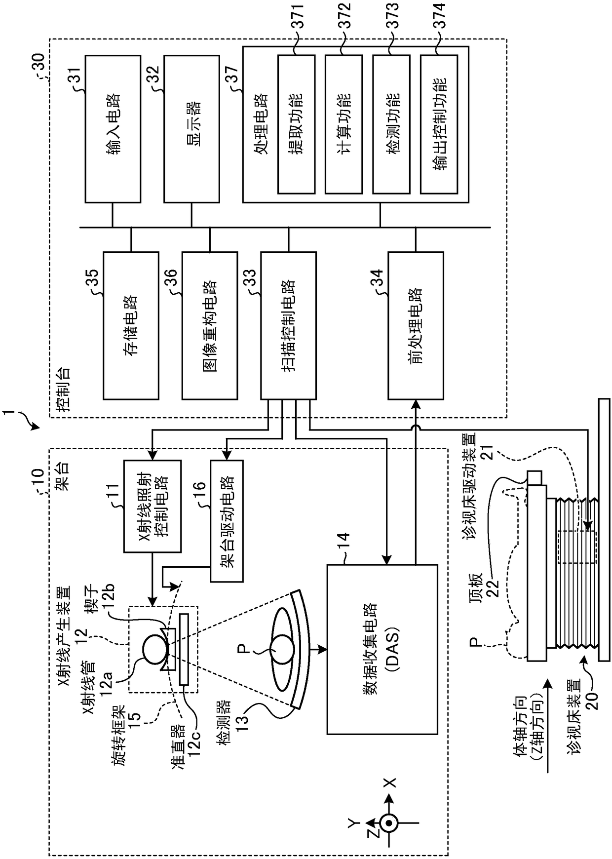
[0026] figure 1 It is a diagram showing an example of the configuration of the X-ray CT apparatus 1 according to the first embodiment. like figure 1 As shown, the X-ray CT apparatus 1 of the first embodiment includes a gantry 10 , a bed apparatus 20 , and a console 30 .

[0027] The gantry 10 is a device that irradiates an object P (patient) with X-rays, detects the X-rays transmitted through the object P, and outputs them to the console 30, and has an X-ray irradiation control circuit 11, an X-ray generator 12, a detection A device 13 , a data acquisition circuit (DAS: Data Acquisition System) 14 , a rotating frame (frame) 15 and a stage driving circuit 16 .

[0028] The rotating frame 15 supports the X-ray generator 12 and the detector 13 so as to face each other across the subject P, and rotates at a high speed on a circular orbit centered on the subject P by a gantry drive circuit 16 described later. circular frame.

[0029] The X-ray irradiation control circuit 11 is ...

no. 2 Embodiment approach

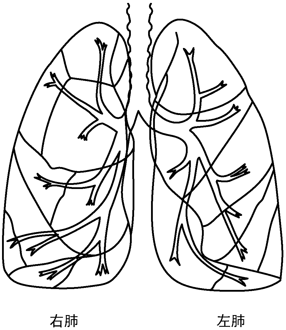
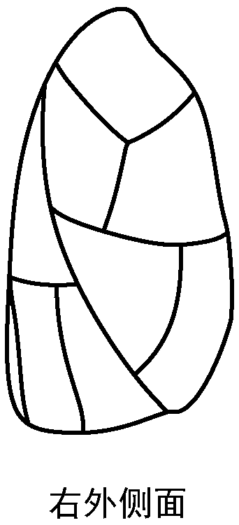
[0114] In the first embodiment, a case was described in which the X-ray CT apparatus 1 detects a region corresponding to a lung lobe or a subregion where an abnormality exists, but the embodiment is not limited thereto. For example, the X-ray CT apparatus 1 can also perform a process of detecting an abnormal region in the bronchi that supplies air to the lung lobes and subregions.

[0115] The X-ray CT apparatus 1 of the second embodiment has the same figure 1 The illustrated X-ray CT apparatus 1 has the same configuration, but part of the processing of the processing circuit 37 is different. Therefore, in the second embodiment, the description will focus on points that are different from the first embodiment, and the description of points that have the same functions as those already described in the first embodiment will be omitted.

[0116] For example, the extraction function 371 further extracts a plurality of bronchi regions corresponding to bronchi that supply air to e...


PUM


Abstract

The embodiment provides a medical image processing apparatus, an X-ray CT apparatus, and a medical image processing method capable of detecting a region of a lung having abnormal breathing function with high precision. The medical image processing apparatus of embodiments includes processing circuitry. The processing circuitry extracts plural regions corresponding to at least one of a lobe and a subsegment forming a lung from three-dimensional medical-image data that is acquired by imaging in temporal order. The processing circuitry calculates a physical index relating to respiratory activityfor each of the extracted regions. The processing circuitry detects an abnormal region having an abnormality relating to the respiratory activity out of the regions by comparing temporal changes of the physical index of the respective regions. The processing circuitry outputs information indicating about the abnormal region.

Description

[0001] References for related applications [0002] This application enjoys the benefits of the priority of Japanese Patent Application No. 2017-017849 filed on February 2, 2017, and the entire content of the Japanese Patent Application is incorporated in this application. In addition, the entire contents of Japanese Patent Application No. 2018-015731 filed on January 31, 2018 are incorporated herein by reference. technical field [0003] Embodiments relate to a medical image processing device, an X-ray CT device, and a medical image processing method. Background technique [0004] Conventionally, in the diagnosis of pulmonary diseases, a process of evaluating respiratory function has been performed. For example, chronic obstructive pulmonary disease (Chronic Obstructive Pulmonary Disease: COPD) and the like are diagnosed by observing the ventilation volume and ventilation curve (spiritual capacity curve) of the entire lung using a spirometer (spirometry). However, for ear...

Claims


Application Information

Patent Timeline
IPC IPC(8): A61B6/03
CPCA61B6/032A61B6/4411A61B6/5217G06T2207/10016G06T11/008G06T2211/412A61B6/488A61B6/5205A61B6/5288A61B6/50A61B6/463G06T2207/30061A61B6/466G06T2207/10081G06T7/0016G16H50/30A61B5/0806G06T7/11G06T7/174G06T11/003
Inventor 五十岚匠真
Owner CANON MEDICAL SYST COPRPORATION
Why Eureka
Try Eureka