Eureka AIR delivers breakthrough ideas for toughest innovation challenges, trusted by R&D personnel around the world.

Bioluminescent detection probe based on transcriptional activator like effector factor and construction method and application thereof

A technology of transcription activation and effector, applied in the field of biomedical analysis, can solve the problems of cumbersome operation, decreased luciferase stability and optical performance, and high price, and achieve the effect of high-sensitivity detection

Active Publication Date: 2018-11-13
SOUTH CENTRAL UNIVERSITY FOR NATIONALITIES
View PDF4 Cites 4 Cited by
  • Summary
  • Abstract
  • Description
  • Claims
  • Application Information

AI Technical Summary

Problems solved by technology

The above chemical labeling process is not only cumbersome and expensive, but also easily leads to the decline of luciferase stability and optical properties.

Method used


Image

  • Bioluminescent detection probe based on transcriptional activator like effector factor and construction method and application thereof
  • Bioluminescent detection probe based on transcriptional activator like effector factor and construction method and application thereof
  • Bioluminescent detection probe based on transcriptional activator like effector factor and construction method and application thereof

Examples


Embodiment 1

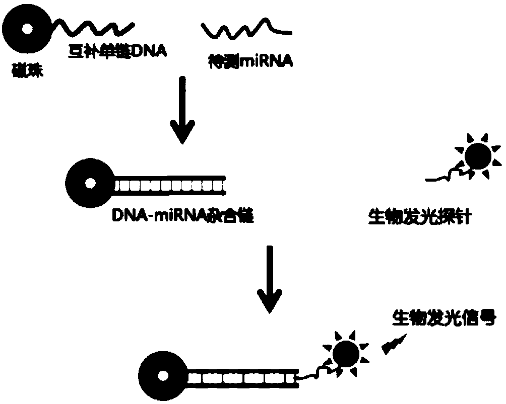
[0028] Example 1: In this example, the miR-21 sequence is used as the object to be detected, and the detection principle is as follows figure 1 shown. In this example, a bioluminescent probe composed of a transcriptional activator-like effector and a luciferase domain is first constructed, and the method is as follows:

[0029] (1) Design and synthesis of the coding fragments of each functional element of the probe: According to the codon preference of E. The coding fragment of the tandem domain and the coding sequence of the luciferase domain; the above coding fragment is prepared by the whole gene synthesis technology, and inserted into the pUC57 plasmid vector.

[0030] (2) Amplification and fusion of the coding fragments of each functional element of the probe: amplify the N-terminal and C-terminal domain coding fragments of transcriptional activator-like effectors by primers P1 and P2, and amplify the fragments at both ends of the amplified product Add the NdeI restrict...

Embodiment 2

[0036] Example 2: Application of a microRNA bioluminescence detection technology based on transcriptional activator-like effectors

[0037] (1) Preparation of ssDNA-coupled magnetic beads: 10 μM biotinylated ssDNA and 1 mg streptavidin-modified magnetic beads were mixed in 50 μL of 1×binding buffer (10 mM Tris-Cl, 1 mM EDTA , 2M NaCl, pH=7.5) for 4h; use a magnetic stand to separate the above magnetic beads, and use 200μL of 1×TALE buffer (20mM Tris-Cl, 60mM KCl, 5mM MgCl 2 , 2mM EDTA, 5% (v / v) glycerol, pH = 8.8) wash the magnetic beads 3 times, resuspend the magnetic beads with 100 μL 5% (w / v) BSA, and place them in a 4°C refrigerator overnight; The blocked magnetic beads were separated with a magnetic stand, resuspended with 100 μL of 1×TALEbuffer, and stored in a 4°C refrigerator until use. The biotinylated single-stranded DNA is complementary to miR-21, and the end is labeled with biotin, and its nucleotide sequence is shown in SEQ ID NO.33; the particles of the magnetic...


PUM


Abstract

The invention provides a bioluminescent detection probe based on a transcriptional activator like effector. The probe can achieve bioluminescence detection of microRNA (miRNA) without chemical labeling. The probe is a monomeric protein, and includes the following three parts: 1) a transcriptional activator like effector domain capable of specifically recognizing a DNA-miRNA hybrid chain; 2) a luciferase domain with bioluminescence; 3) a connecting region of the above two domains. The probe uses a single-stranded DNA coupled to the surface of a magnetic bead to hybridize with the miRNA to be tested, and a DNA-miRNA hybrid chain produced by hybridization is specifically recognized by the bioluminescent probe formed by the transcriptional activator like effector and the luciferase domain to achieve high sensitivity detection of the miRNA. The probe has the advantages of being low in cost, intuitive in result and convenient to use.

Description

technical field [0001] The invention relates to a microRNA bioluminescent detection technology based on a transcriptional activator-like effector, belonging to the field of biomedical analysis. Background technique [0002] MicroRNA (miRNA) is an important biomarker, and its abnormal expression and metastasis in the human body are closely related to the occurrence and development of major diseases such as tumors. Therefore, the high-sensitivity detection method of miRNA has broad market prospects in the fields of early diagnosis of diseases, research on pathogenesis and related drug development. [0003] In recent years, bioluminescent technology has been applied in miRNA detection due to its advantages of high sensitivity and specificity. This technology uses luciferase-catalyzed substrate luminescence as the detection signal and does not require external light source excitation, so it can effectively avoid the disadvantages of biological autofluorescence interference, pho...

Claims


Application Information

Patent Timeline
IPC IPC(8): C12Q1/6816C12Q1/66
CPCC12Q1/66C12Q1/6816C12Q2525/207C12Q2563/143C12Q2563/149
Inventor 李勇吴云华
Owner SOUTH CENTRAL UNIVERSITY FOR NATIONALITIES
Why Eureka
Eureka Blog
Learn More