A method for establishing an animal model of retinal edema

A retinal edema and animal model technology, applied in medical science, veterinary instruments, animal husbandry, etc., can solve the problems of retinal atrophy, vascular damage to the retina, and increased retinal damage, achieving small trauma and good repeatability

Active Publication Date: 2022-02-15
SHANDONG UNIV QILU HOSPITAL
View PDF4 Cites 0 Cited by
  • Summary
  • Abstract
  • Description
  • Claims
  • Application Information

AI Technical Summary

Problems solved by technology

Some scholars prepared a retinal edema model by occluding retinal veins: using SD rats, intravenously injecting photosensitizer Erythrosin B combined with 532nm laser to continuously irradiate the retina for 8 minutes, blood vessel damage and retinal thickness increased immediately after operation, but retinal thickness increased 5 days after operation. Recovered to pre-operation, retinal atrophy occurred 14 days later, unable to provide an animal model of long-term retinal edema, and long-term laser irradiation increased damage to other parts of the retina

Method used


Image

  • A method for establishing an animal model of retinal edema
  • A method for establishing an animal model of retinal edema
  • A method for establishing an animal model of retinal edema

Examples


Embodiment 1

[0046] Example 1: Establishment of retinal edema model using BN rats

[0047] Step 1: Preparation of Rose Bengal Solution

[0048] Under the condition of avoiding light, accurately weigh 50 mg of Rose Bengal powder, place it in a 1.5 ml brown EP tube, add 1.0 ml of normal saline, and mix well. After filtering three times with a 0.22 μm sterile filter, the prepared Rose Bengal solution (50 mg / ml) was stored at 4° C. for use.

[0049] Step 2: BN rat preparation

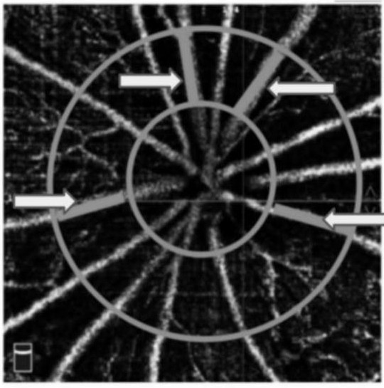
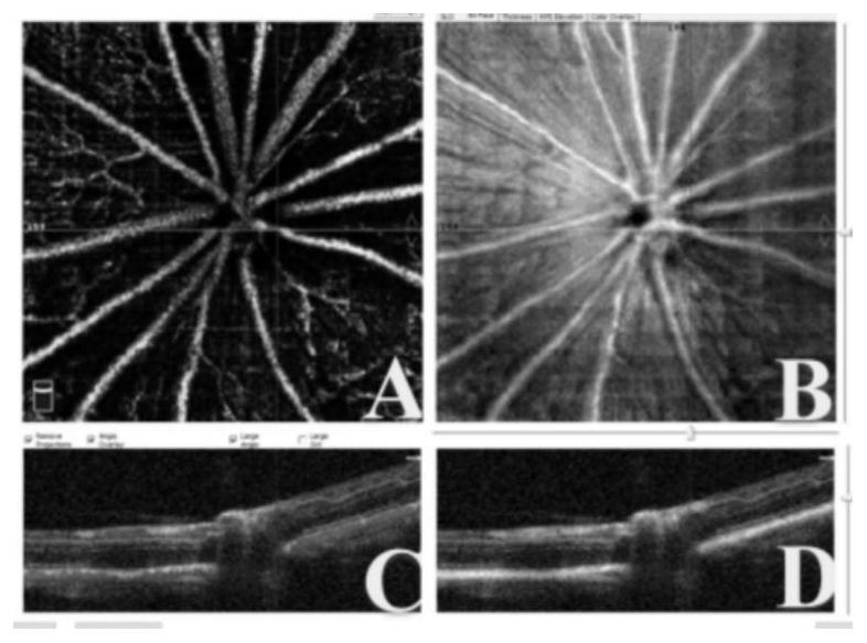
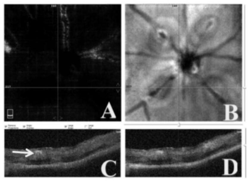
[0050] Four female BN rats, aged 7-8 weeks, with an average body weight of 150 g, were intraperitoneally injected with 10% (w / w) chloral hydrate (0.3 ml / 100 g). After anesthesia, compound tropicamide eye drops fully dilated mydriasis, slit lamp, OCTA / OCT examination, records ( figure 2 ). Fix the rats and fully disinfect the tail with 75% alcohol. 50mg / ml Rose Bengal solution (0.1ml / 100g) was left to stand at room temperature for 10 minutes and then injected into the tail vein of rats. After the injection, it was ...

Embodiment 2

[0058] Example 2: Establishment of retinal edema model using C57BL / 6 mice

[0059] Step 1: Preparation of Rose Bengal Solution

[0060] Under dark conditions, accurately weigh 25 mg of Rose Bengal powder, place it in a 1.5 ml brown EP tube, add 1.0 ml of normal saline, and mix well. After filtering three times with a 0.22 μm sterile filter, a Rose Bengal solution (25 mg / ml) was prepared and stored at 4° C. for future use.

[0061] Step 2: C57BL / 6 mouse preparation

[0062] 15 female C57BL / 6 mice, 6-8 weeks old, with an average body weight of 20g, were injected intraperitoneally with 10% (w / w) chloral hydrate (1ml / 100g), and after anesthesia, compound tropicamide eye drops were fully administered. Mydriasis, slit lamp, OCTA and OCT examination, records. Fix the mouse and fully disinfect the tail with 75% alcohol. 25mg / ml Rose Bengal solution (0.5ml / 100g) was left to stand at room temperature for 10 minutes and then injected into the rat tail vein. After the injection, it wa...


PUM


Abstract

The invention discloses a method for establishing an animal model of retinal edema. The rational application of 577nm laser combined with Rose Bengal was used to occlude retinal veins in mice to form retinal edema. Non-invasive OCTA / OCT technology was used to continuously observe the retina of the model eye and its edema state. The present invention is devoted to inventing a simple and easy-to-use method for establishing a retinal edema animal model with little trauma to the model animal, fast onset of effect, long maintenance time, so as to well meet the corresponding experimental requirements.

Description

technical field [0001] The invention relates to a method for establishing an animal model of retinal edema. Background technique [0002] As a common eye disease, macular edema is more common in retinal vein occlusion, diabetic retinopathy, chronic uveitis, ocular trauma, and after intraocular surgery. The patients felt that their visual acuity decreased and their metamorphopsia gradually worsened, which seriously affected the quality of life of the patients. The pathogenesis of macular edema is mainly due to the leakage of capillary damage in the macular area caused by the above-mentioned primary diseases, resulting in edema. At present, a large number of scientific researchers are committed to the development of drugs for the treatment of macular edema. Preclinical experiments require an animal model of retinal edema to observe the therapeutic effect of the drug being developed. The establishment of the model requires simplicity, low trauma, quick onset and long mainten...

Claims


Application Information

Patent Timeline
Patent Type & Authority Patents(China)
IPC IPC(8): A01K67/02A61D7/00
CPCA01K67/02A61D7/00
Inventor 曲毅程同杰程英李靳
Owner SHANDONG UNIV QILU HOSPITAL
Why Eureka
Try Eureka