Visible ureteroscope dilatation balloon and use method thereof

A ureteroscope and balloon technology, applied in ureteroscope, application, endoscope and other directions, can solve the problems of ureteral injury, long operation time, inaccurate expansion position, etc., and achieve the effect of ensuring support performance

Pending Publication Date: 2018-12-07
苏州市倍咏医疗科技有限公司
View PDF10 Cites 4 Cited by
  • Summary
  • Abstract
  • Description
  • Claims
  • Application Information

AI Technical Summary

Problems solved by technology

[0002] Rigid ureteroscope dilation is currently the mainstream method. However, since the rigid mirror is made of metal and has high hardness, direct expansion may cause complications such as ureteral injury, ureteral tear, and perforation. However, there are many models and specifications of ureteral dilators. Step-by-step dilation may be required, and the dilation cannot be directly viewed under the endoscope, which may lead to inaccurate dilation positions, which in turn lead to complications such as normal ureteral injury, and the operation time is longer, while the existing balloon dilation is currently widely used For intravascular stenosis, gastrointestinal stenosis and other departments, balloon dilatation has the advantages of not requiring step-by-step expansion, the expansion pressure can be adjusted according to the degree of stenosis, and the expansion process is relatively gentle, which can solve the problem of long operation time , but it also has the same problem as the ureteral dilator. The working hole of the current flexible ureteroscope is only 1.2mm, and the outer diameter of the balloon after folding reaches 1-1.5mm, that is, it cannot be expanded under the direct vision of the endoscope. Dilation under X-ray positioning, because dilation cannot be performed under direct vision, may cause ureteral perforation, rupture, etc. The operation is difficult and the operation effect is not very satisfactory

Method used


Image

  • Visible ureteroscope dilatation balloon and use method thereof
  • Visible ureteroscope dilatation balloon and use method thereof
  • Visible ureteroscope dilatation balloon and use method thereof

Examples


Embodiment 1

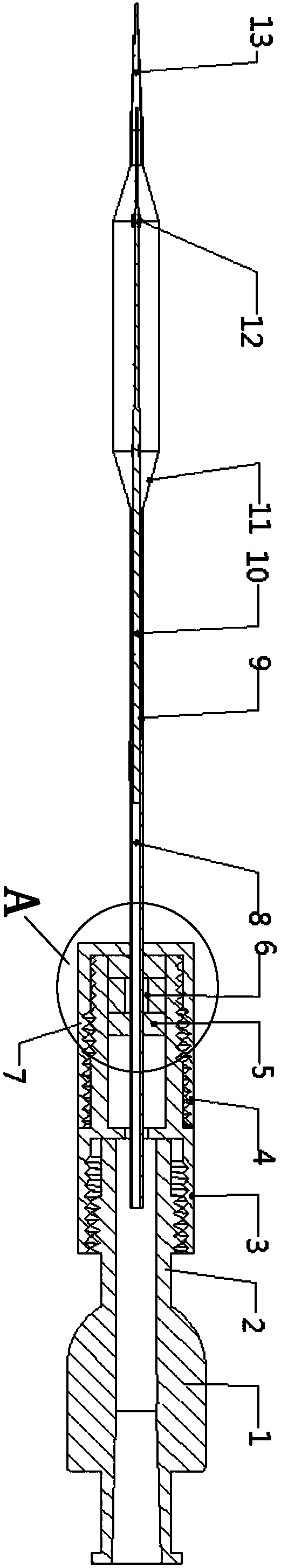
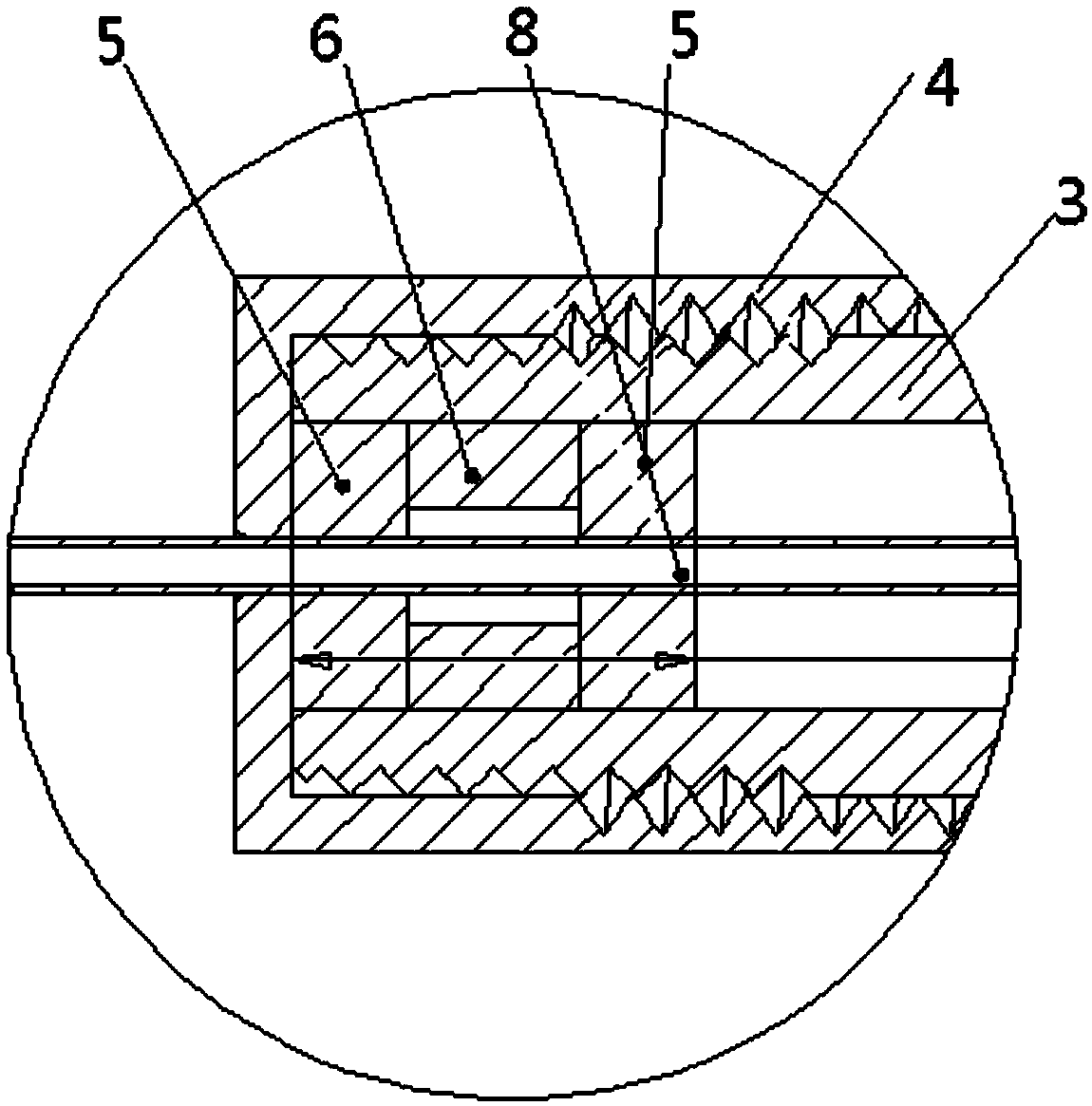
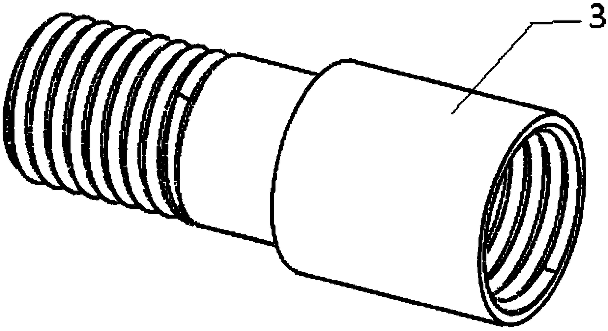
[0029] Such as Figure 1-3 The shown dilatation balloon of the present invention for a visual ureteroscope includes a joint assembly 1, an air guiding channel is arranged inside the joint assembly 1, and a rigid catheter 8 is fixed at one end of the joint assembly 1 detachably. One end of the hard catheter 8 communicates with the air guide channel, the other end of the hard catheter 8 communicates with a soft catheter 9, and the soft catheter 9 is provided with a balloon 11. When the present invention is in use, , connected to the joint assembly 1 through a pressure pump, the airflow passes through the air guide channel, the hard catheter 8 and the soft catheter 9 to pressurize the balloon 11, and the head of the balloon 11 is provided with a soft guide 13, so The soft catheter 9 is used to be inserted into the urethra, so nylon or other modified polymers are used to prevent damage to human body components. The soft catheter 9, the balloon 11 and the soft guide head 13 are pro...


PUM


Abstract

The invention relates to a visible ureteroscope dilatation balloon and a use method thereof. A joint assembly is internally provided with a gas guiding passage, a hard catheter is detachably and fixedly clamped to one end of the joint assembly, one end of the hard catheter is communicated with the gas guiding passage, the other end of the hard catheter is communicated with a soft catheter, the soft catheter is provided with a balloon body, the head of the balloon body is provided with a soft guiding head, and a guiding wire penetrates through the soft catheter, the balloon body and the soft guiding head. According to the visible ureteroscope dilatation balloon and the use method thereof, through the arrangement of the hard catheter and the joint assembly detechably and fixedly clamped to the hard catheter, the hard catheter can be detached and inserted from the far end of a flexible ureteroscope, penetrates out of the near end and is fixedly clamped to the joint assembly, the dilatation balloon and the flexible ureteroscope are used together, and the situation is avoided that in the prior art, the ureteroscope is used for examination at first and then the dilatation balloon is usedfor dilatation, so that positioning is inaccurate, and perforation or rupture of ureters is caused.

Description

technical field [0001] The invention relates to a visual ureteroscope dilatation balloon and a method for using the same, belonging to the technical field of medical equipment. Background technique [0002] Rigid ureteroscope dilation is currently the mainstream method. However, since the rigid mirror is made of metal and has high hardness, direct expansion may cause complications such as ureteral injury, ureteral tear, and perforation. However, there are many models and specifications of ureteral dilators. Step-by-step dilation may be required, and the dilation cannot be directly viewed under the endoscope, which may lead to inaccurate dilation positions, which in turn lead to complications such as normal ureteral injury, and the operation time is longer, while the existing balloon dilation is currently widely used For intravascular stenosis, gastrointestinal stenosis and other departments, balloon dilatation has the advantages of not requiring step-by-step expansion, the e...

Claims


Application Information

Patent Timeline
Patent Type & Authority Applications(China)
IPC IPC(8): A61B1/32A61B1/307
CPCA61B1/00071A61B1/307A61B1/32
Inventor 徐星岗常健王乐康
Owner 苏州市倍咏医疗科技有限公司
Why Eureka
Try Eureka