Periodontal Endoscope with Disposable Cannula and 3D Steering

An endoscope, a disposable technology, applied in the field of endoscopy, can solve the problems of prolonged dental operation time, blurred images, inconvenience, etc., and achieve the effects of convenient clinical application, reduced volume, and reliable combination

Active Publication Date: 2020-11-24
陕西飞秒医疗设备有限公司
View PDF3 Cites 0 Cited by
  • Summary
  • Abstract
  • Description
  • Claims
  • Application Information

AI Technical Summary

Problems solved by technology

The main disadvantage of this type of endoscope is that the objective lens is located at the front end of the periodontal endoscope. After the objective lens enters the periodontal cavity, it is blocked by tissue fluid, and the image displayed on the display screen is blurred, and the lesion condition of the periodontal cavity cannot be clearly seen.
Moreover, in its structure, the endoscope is always in contact with the human body. Due to the sophisticated optical components and image sensors in the structure of the endoscope, it cannot withstand repeated high-temperature and high-pressure cooking sterilization methods for a long time. The sterilization method does not meet the sterilization requirements of dental surgery, and the endoscope is in direct contact with human tissue and blood, resulting in a significant increase in the risk of cross-infection
[0004] The handle of the existing periodontal endoscope has only one fixed angle, and the angle cannot be adjusted during use, so that the endoscope can only perform dental surgery in one direction. During the operation, the operator needs to replace the handle with a different angle to complete the operation. Dental surgery, which is inconvenient and prolongs the time of dental surgery

Method used


Image

  • Periodontal Endoscope with Disposable Cannula and 3D Steering
  • Periodontal Endoscope with Disposable Cannula and 3D Steering
  • Periodontal Endoscope with Disposable Cannula and 3D Steering

Examples


Embodiment Construction

[0030] In order to make the object, technical solution and advantages of the present invention clearer, the present invention will be further described in detail below in conjunction with the accompanying drawings and embodiments. It should be understood that the specific embodiments described here are only used to explain the present invention, not to limit the present invention.

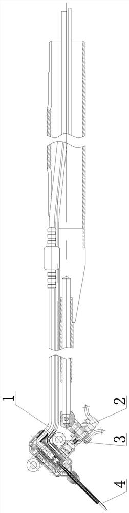
[0031] exist figure 1 Among them, the periodontal endoscope with a disposable sleeve 3 in this embodiment is composed of an endoscope 1, a handle 2, a disposable sleeve 3, and an expansion tube 4.

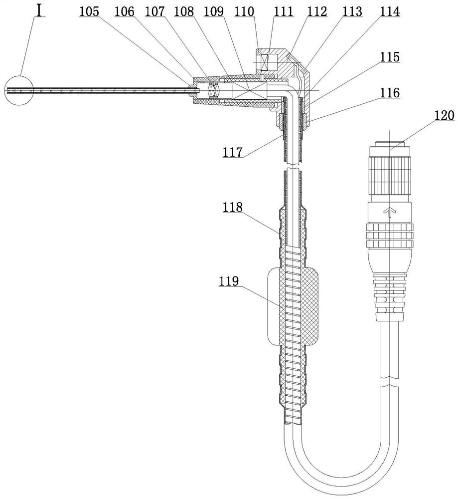
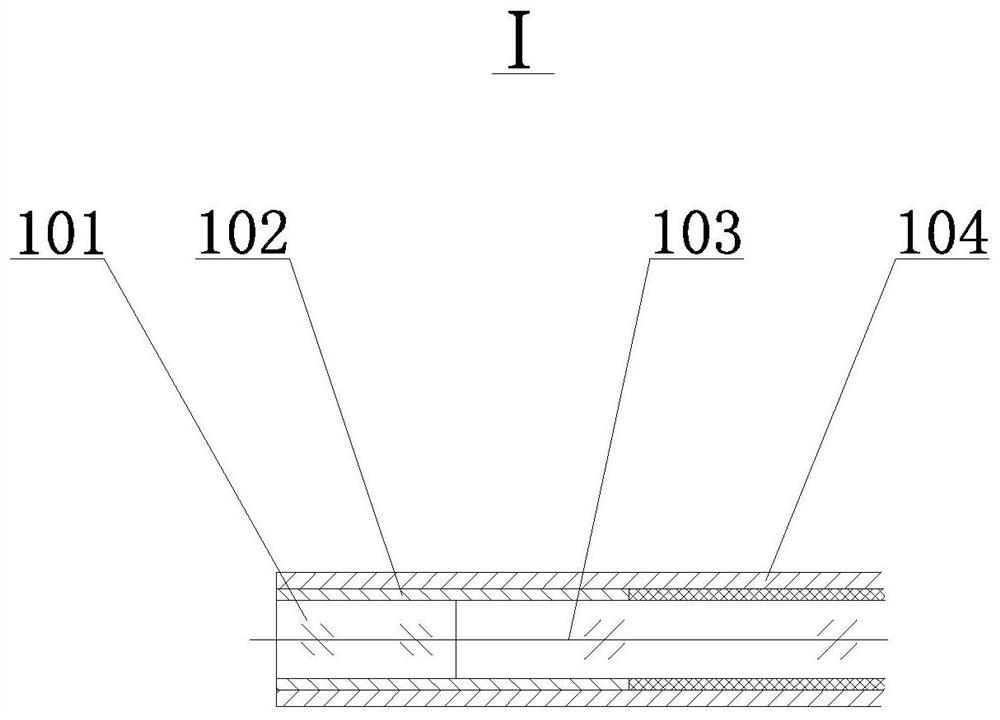
[0032] exist figure 2 , 3 Among them, the endoscope 1 of present implementation is made of objective lens 101, objective lens barrel 102, image-passing rod 103, mirror tube 104, coupling lens barrel 105, mirror cone 106, coupling lens 107, camera cover 108, camera 109, gland 110 , LED light-emitting diode 111, mirror body base 112, cable gland 113, flexible spring tube 114, cable clamp 115, pull rin...


PUM


Abstract

The invention relates to a three-dimensional-steering periodontal endoscope with a disposable sleeve. The periodontal endoscope comprises an endoscope body, a handle, the disposable sleeve and an expansion tube and is characterized in that the handle is arranged on the endoscope body, the disposable sleeve is arranged on the endoscope body, and the expansion tube is arranged on the handle; a camera and a camera sleeve are mounted in an endoscope cone transversely arranged on an endoscope body base, a coupling lens is arranged on the left side of the camera in the camera sleeve, the endoscope cone is provided with a mirror tube with an object slide, an objective lens and an image transmitting rod are arranged on the left side in the object slide, the upper portion of the endoscope body baseis provided with an LED, and the right side of the endoscope body base is provided with a cable-passing gland; the endoscope body base is vertically provided with a flexible spring tube, a cable clamp is arranged outside one end of the flexible spring tube, the other end of the flexible spring tube is provided with a standard connector, a rubber sleeve is arranged outside the upper portion of theflexible spring tube, and the lower portion of the flexible spring tube is provided with cable clamp sleeve with a cable jacket outside. The periodontal endoscope is reasonable in design, small in size, multifunctional, convenient to use, clear in image display, capable of being used as a periodontal diagnosis and surgery instrument and the like.

Description

technical field [0001] The invention belongs to the technical field of endoscopes, in particular to periodontal endoscopes. Background technique [0002] Periodontal disease is relatively common. For gingivitis and mild and moderate periodontitis, it can be treated by brushing teeth, using dental floss and cleaning the mouth. Moderate and severe periodontitis is more complicated. Reversal, the main means of treatment is to control inflammation and prevent further development of inflammation. The commonly used methods are scaling and root planing. The periodontal is cut open with a scalpel and the flap is turned to expose the calculus on the subgingival root surface. After the flap is turned, the calculus is removed under the condition of direct vision. This operation method has the disadvantages of heavy damage to the patient, great pain, long postoperative healing time and inability to eat well. [0003] In order to solve the above-mentioned technical problems in the trea...

Claims


Application Information

Patent Timeline
Patent Type & Authority Patents(China)
IPC IPC(8): A61B1/247A61B1/06A61B1/12A61B1/05
CPCA61B1/00114A61B1/05A61B1/0684A61B1/12A61B1/247
Inventor 李小刚骆永明常婧媛万燕玲陈鹏
Owner 陕西飞秒医疗设备有限公司
Why Eureka
Try Eureka