Method for preparing micro-nanostructure sample by using focused ion beam scanning electron microscope double beam system

A technology of focusing ion beam and scanning electron microscope, which is applied in the preparation of nanostructured samples and in the micro field, can solve the problems of difficulty in the growth of micro and nanostructures, and achieve the effect of improving real-time performance and reducing costs.

Inactive Publication Date: 2018-12-11
HEFEI INSTITUTES OF PHYSICAL SCIENCE - CHINESE ACAD OF SCI
View PDF8 Cites 3 Cited by
  • Summary
  • Abstract
  • Description
  • Claims
  • Application Information

AI Technical Summary

Problems solved by technology

Although this method of using a dual-beam system to prepare samples with micro-nano structures can obtain samples of various shapes, there is also a need for micro-na...

Method used


Image

  • Method for preparing micro-nanostructure sample by using focused ion beam scanning electron microscope double beam system
  • Method for preparing micro-nanostructure sample by using focused ion beam scanning electron microscope double beam system
  • Method for preparing micro-nanostructure sample by using focused ion beam scanning electron microscope double beam system

Examples


Embodiment 1

[0035] The concrete steps of preparation are:

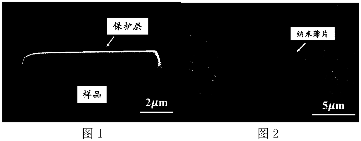
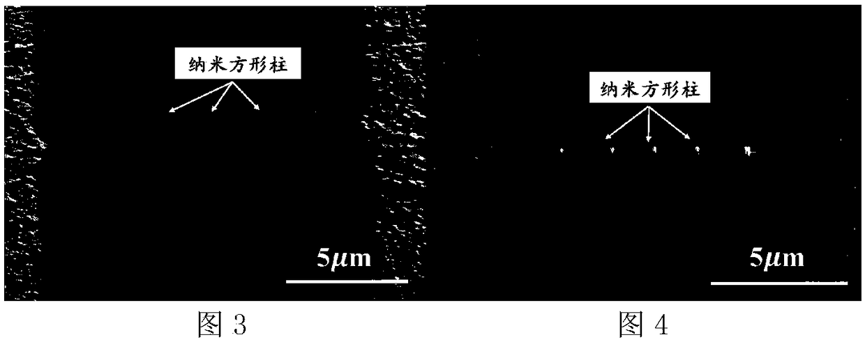
[0036] Step 1, prior to the surface deposition of the sample than the edge of the area to be measured 500nm wider, thick 0.5 μm containing organic protection layer; wherein, the deposition is to use electron beam deposition (or use ion beam deposition), the organic protection layer is Gold layer with organics. Then the sample is cut into micro-nano flakes with the same width as the protective layer.

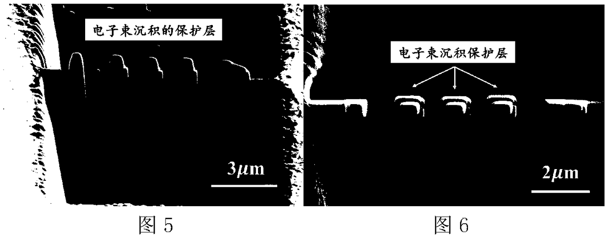
[0037] In step 2, the micro-nano sheet is first cut into columns whose cross-section is the shape to be measured; wherein, the shape to be measured is a rectangle which is one of the nano-polygons. After cleaning the amorphous layer on the surface of the columnar surface with an ion beam, use an electron beam to deposit a protective layer containing organic matter with a thickness of 50nm and an ion beam deposition thickness of 2μm; wherein, the protective layer containing organic matter is a gold layer containing organic matter ....

Embodiment 2

[0040] The concrete steps of preparation are:

[0041] Step 1, prior to the surface deposition of the sample than the edge of the area to be measured 600nm wider, thick 0.88 μm containing organic protective layer; wherein, the deposition is to use electron beam deposition (or use ion beam deposition), the protective layer containing organic matter is Gold layer with organics. Then the sample is cut into micro-nano flakes with the same width as the protective layer.

[0042] In step 2, the micro-nano sheet is first cut into columns whose cross-section is the shape to be measured; wherein, the shape to be measured is a rectangle which is one of the nano-polygons. After cleaning the amorphous layer on the surface of the columnar surface with an ion beam, use an electron beam to deposit a protective layer with a thickness of 163 nm and an ion beam with a thickness of 1.8 μm. layer.

[0043] Step 3, first use an ion beam to cut the columnar sample covered with an organic-contain...

Embodiment 3

[0045] The concrete steps of preparation are:

[0046] Step 1, prior to the surface deposition of the sample than the edge of the area to be measured 700nm wider, thick 1.25 μm containing organic protection layer; wherein, the deposition is to use electron beam deposition (or use ion beam deposition), the organic protection layer is Gold layer with organics. Then the sample is cut into micro-nano flakes with the same width as the protective layer.

[0047]In step 2, the micro-nano sheet is first cut into columns whose cross-section is the shape to be measured; wherein, the shape to be measured is a rectangle which is one of the nano-polygons. After cleaning the amorphous layer on the columnar surface with an ion beam, use an electron beam to deposit a protective layer containing organic matter with a thickness of 273nm and an ion beam deposition thickness of 1.5 μm; wherein, the protective layer containing organic matter is gold containing organic matter layer.

[0048] Ste...


PUM


Abstract

The invention discloses a method for preparing a micro-nanostructure sample by using a focused ion beam scanning electron microscope double beam system. The method comprises the following steps of firstly, depositing an organic matter contained protective layer with the edge wider than the edge of a region to be detected on the surface of a sample; then cutting the sample into a micro-nano thin slice with the same width as the protective layer; then cutting the micro-nano thin slice into a column shape with the cross section being a morphology to be detected, then depositing the organic mattercontained protective layer on the column-shaped surface by sequentially using an electron beam and an ion beam; and finally, cutting the column-shaped sample, the surface of which is covered with theorganic matter contained protective layer, into a thin slice along the cross section by using the ion beam firstly, and then cleaning an amorphous layer on the surface of the thin slice by using theion beam to prepare the micro-nanostructure sample. The method can be rapidly and effectively used in a bulk material to process the samples in required morphologies and sizes, as samples of a scanning electron microscope, a transmission electron microscope, electric transport measurement, ferromagnetic resonance test and the like, and is a universal method for manufacturing the micro-nanostructure samples in various morphologies.

Description

technical field [0001] The invention relates to a method for preparing a micro-nano structure sample, in particular to a method for preparing a micro-nano structure sample by using a focused ion beam scanning electron microscope dual-beam system. Background technique [0002] The focused ion beam-electron beam system is a system that combines the ion beam and the electron beam. This system combines the cutting function of the high-energy ion beam with the imaging function of the electron beam, thereby realizing the cutting of the sample and the image of the sample at the same time. Real-time observation; In addition, with the assistance of organic chemical gases, electron beams and ion beams can also achieve the deposition function of special materials. With the gradual maturity of the dual-beam system technology, its application range is becoming wider and wider, and one of the most important aspects is the processing of micro- and nano-structured samples; MnSi nanowires",...

Claims


Application Information

Patent Timeline
IPC IPC(8): G01N23/2202
CPCG01N23/2202
Inventor 杜海峰王莎莎田明亮
Owner HEFEI INSTITUTES OF PHYSICAL SCIENCE - CHINESE ACAD OF SCI
Why Eureka
Try Eureka