MRI image-based axillary lymph gland metastasis prediction system

A technology of lymph node metastasis and prediction system, applied in the field of computer-aided diagnosis, can solve the problems of interference, difficulty in display and diagnosis, poor spatial resolution of axillary lymph nodes, etc., and achieve the effect of improving accuracy and efficiency

Inactive Publication Date: 2018-12-18
NORTHEASTERN UNIV
View PDF5 Cites 20 Cited by
  • Summary
  • Abstract
  • Description
  • Claims
  • Application Information

AI Technical Summary

Problems solved by technology

Because there are structures such as pectoralis major and pectoralis minor around the lymph nodes in zone II, and there is interference from apical air in zone III, it is difficult to display and diagnose these two groups of lymph nodes by ultrasound.
Magnetic resonance imaging is a non-invasive and radiation-free examination metho...

Method used


Image

  • MRI image-based axillary lymph gland metastasis prediction system
  • MRI image-based axillary lymph gland metastasis prediction system
  • MRI image-based axillary lymph gland metastasis prediction system

Examples


Embodiment Construction

[0039] The specific implementation manners of the present invention will be further described in detail below in conjunction with the accompanying drawings and embodiments. The following examples are used to illustrate the present invention, but are not intended to limit the scope of the present invention.

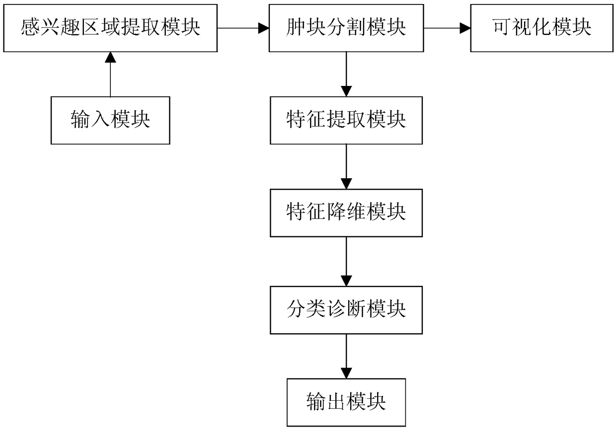
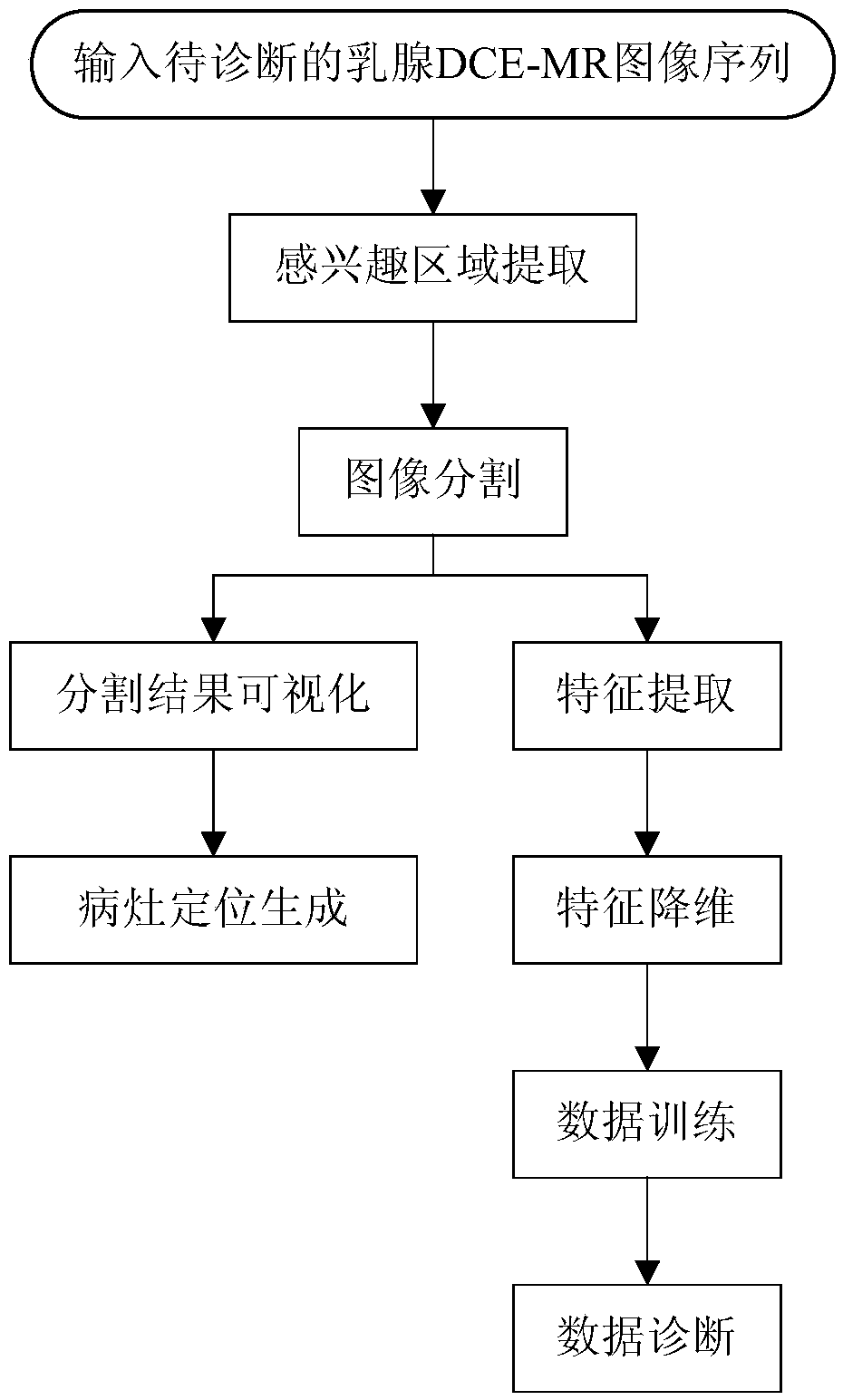
[0040] Accurate prediction of axillary lymph node status is of great significance for breast cancer patients with negative axillary lymph nodes, which can avoid unnecessary axillary lymph node dissection and reduce pain and cost. Such as figure 1 As shown, the axillary lymph node metastasis prediction system based on MRI images provided in this embodiment includes an input module, a region of interest extraction module, a tumor segmentation module, a visualization module, a feature extraction module, a feature dimensionality reduction module, a classification diagnosis module and an output module . The method flow of using the system of this embodiment to predict the met...


PUM


Abstract

The invention provides an MRI image-based axillary lymph gland metastasis prediction system and relates to the technical field of computer aided diagnosis. The system comprises an input module, an area-of-interest extraction module, a lump segmentation module, a sub-visualization module, a feature extraction module, a feature dimensionality reduction module, a classification and diagnosis module,and an output module. The input module receives a to-be-diagnosed mammary gland DCE-MR image sequence input by a user. The area-of-interest extraction module extracts an area of interest from the mammary gland DCE-MR image sequence. The lump segmentation module segments a lump in the area of interest. The sub-visualization module carries out the visual display on each segmented image and extractsthe edge of a focus. The feature extraction module extracts relevant feature values according to the lump information and transmits the relevant feature values to the feature dimensionality reductionmodule. The feature dimensionality reduction module carries out feature dimensionality reduction on an extracted feature set. The classification and diagnosis module inputs each lump feature value into a classifier. After that, the automatic classification and recognition is carried out by a computer for judging whether a lymph gland has already been transferred or not. The output module displaysa transfer prediction result and a transfer probability. According to the invention, the accurate segmentation of breast lesions can be realized. The accurate diagnosis of mammary axillary lymph glandmetastasis can be effectively assisted.

Description

technical field [0001] The invention relates to the technical field of computer-aided diagnosis, in particular to an MRI image-based axillary lymph node metastasis prediction system. Background technique [0002] Breast cancer is the most common malignant tumor in women worldwide and the second leading cause of cancer death in women after lung cancer. Breast cancer cells are likely to metastasize when the diameter of the tumor is greater than 2cm. Breast cancer cells often metastasize to the ipsilateral axillary lymph nodes first, and the greater the number of metastatic lymph nodes, the lower the patient's survival rate. The presence or absence of axillary lymph node metastasis in breast cancer patients is of great significance to the staging, treatment and prognosis of breast cancer, and it is also one of the important reference indicators for postoperative radiotherapy and chemotherapy. [0003] At present, the gold standard for diagnosis of lymph node metastasis is sti...

Claims


Application Information

Patent Timeline
IPC IPC(8): A61B5/055
CPCA61B5/055A61B5/7275
Inventor 赵越王念崔笑宇郑靖巩立鑫
Owner NORTHEASTERN UNIV
Why Eureka
Try Eureka