Intracranial hemorrhage blood clot locating system and method based on CT image
A technology of CT imaging and intracranial hemorrhage, which is applied in the field of blood clot positioning system for intracranial hemorrhage, can solve the problems of lack of reliability and accuracy, damage to brain tissue, etc., and achieve the effect of improving treatment efficiency and good treatment effect
- Summary
- Abstract
- Description
- Claims
- Application Information
AI Technical Summary
Problems solved by technology
Method used
Image

Examples
Embodiment 1
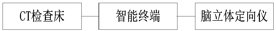
[0041] This embodiment provides a blood clot location system for intracranial hemorrhage based on ct images, such as figure 1 As shown, it includes a CT examination bed, a brain stereotaxic instrument and an intelligent terminal, and the CT examination bed and the brain stereotaxic instrument communicate with the intelligent terminal respectively;
[0042] The CT examination bed is used to scan the brain, and send the scanned CT image data to the smart terminal, and the smart terminal analyzes the CT image to obtain blood clot coordinates, and controls the brain stereotaxic instrument to detect the blood clot according to the blood clot coordinates. Accurate positioning and puncture removal.
[0043] In this embodiment, the blood clot coordinates are obtained by analyzing the CT image data, and the stereotaxic instrument is controlled according to the blood clot coordinates to accurately locate and puncture and remove the blood clot, which improves the treatment efficiency and...
Embodiment 2
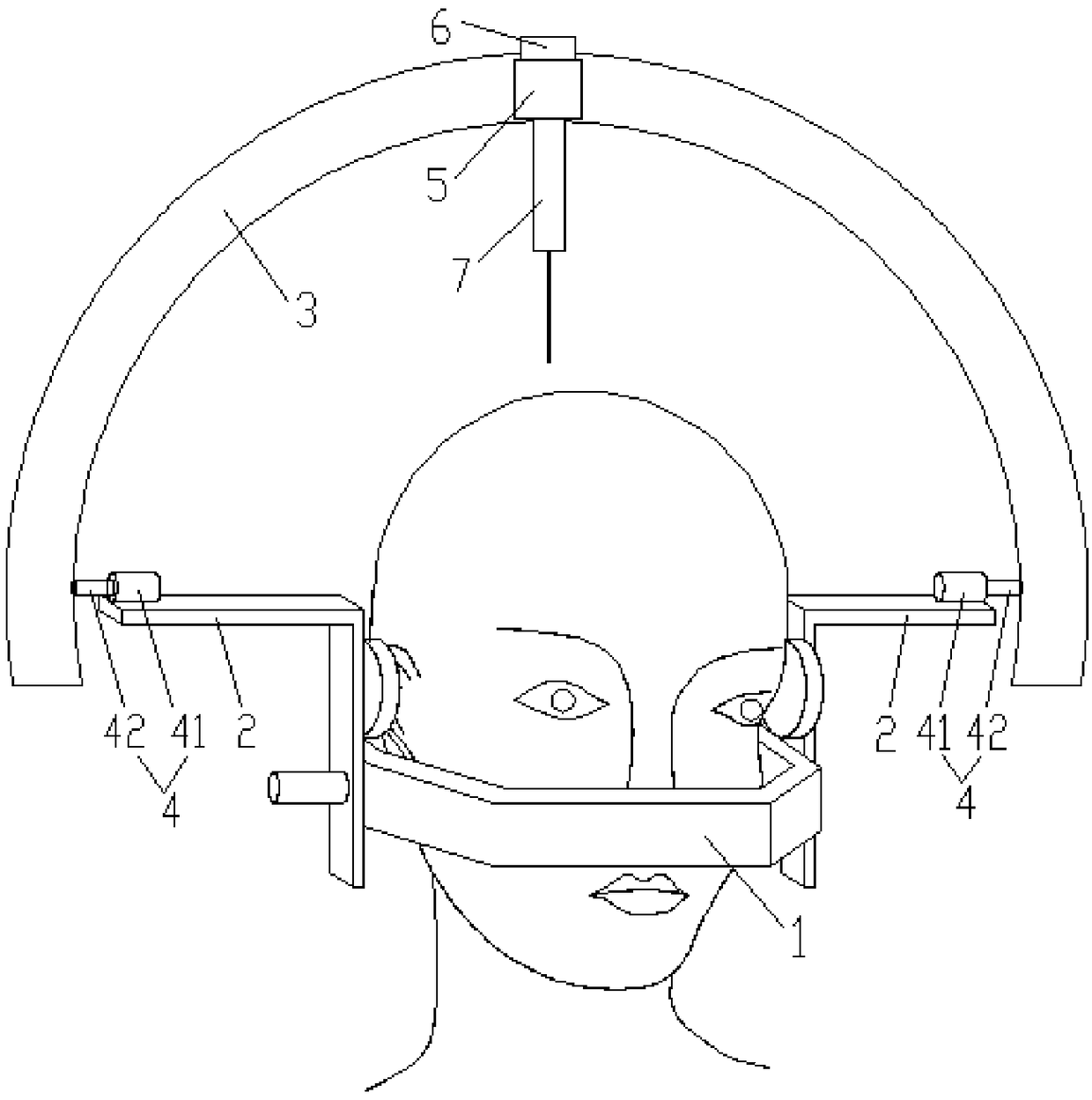
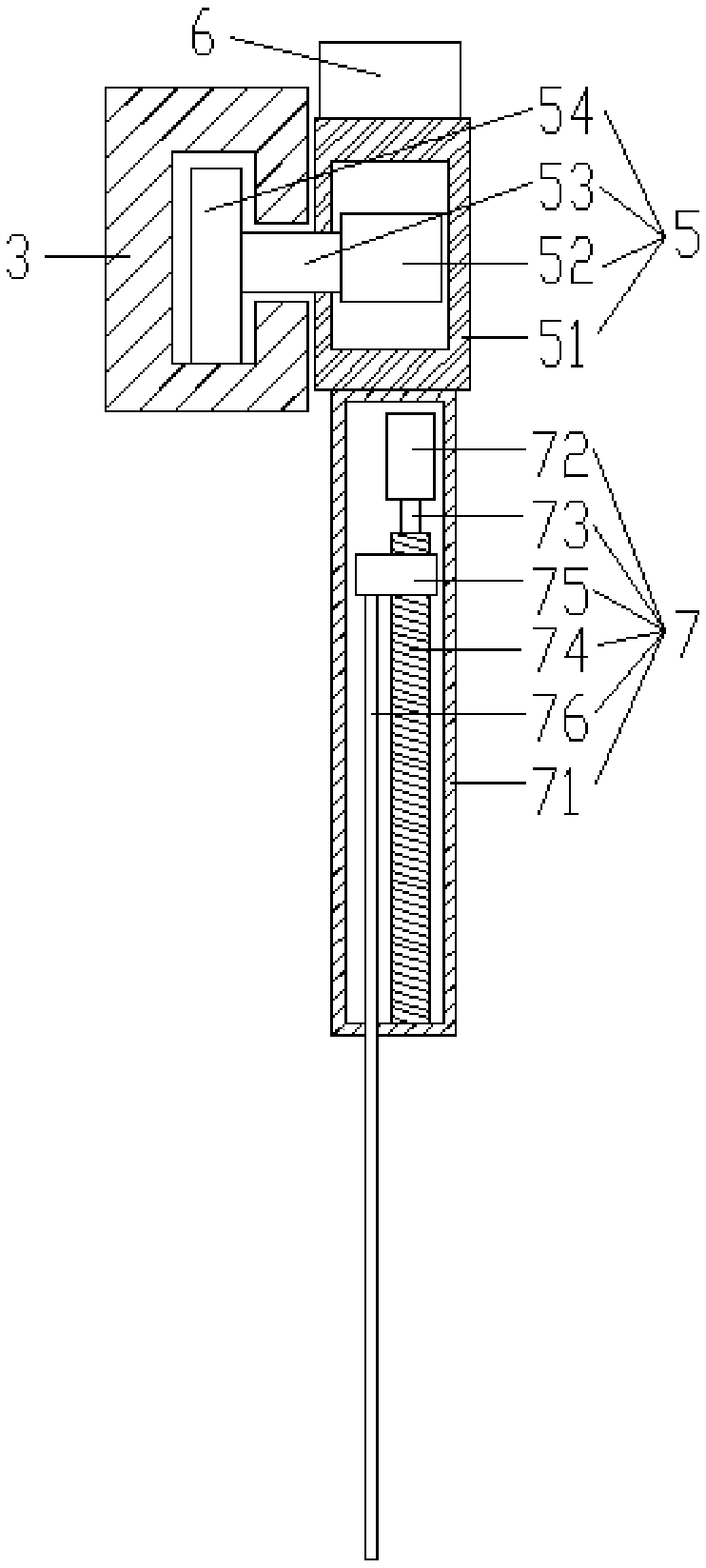
[0045] This embodiment provides a blood clot localization system for intracranial hemorrhage based on CT images, such as Figure 1-Figure 4 As shown, it includes a CT examination table, a brain stereotaxy apparatus and an intelligent terminal, and the CT examination table and the brain stereotaxy apparatus communicate with the intelligent terminal respectively. The CT examination table of this embodiment adopts multi-slice spiral CT. Compared with ordinary CT, multi-slice spiral CT can complete a large range of volume scanning at high speed, with good image quality, fast imaging speed, high longitudinal resolution and excellent time resolution. The smart terminal of this implementation adopts a computer or other smart devices.
[0046] The CT examination bed is used to scan the brain, and send the scanned CT image data to the smart terminal, and the smart terminal analyzes the CT image to obtain blood clot coordinates, and controls the brain stereotaxic instrument to detect t...
Embodiment 3
[0058] This embodiment provides a blood clot location method for intracranial hemorrhage based on CT images, which is applicable to the blood clot location system for intracranial hemorrhage based on CT images described in Embodiment 2, such as Figure 5 shown, including the following steps:
[0059] S1, the CT examination table scans the brain, and sends the scanned CT image data to the smart terminal;
[0060] S2, the intelligent terminal analyzes the CT image to obtain the coordinates of the blood clot, and sends the work order and the coordinates of the blood clot to the stereotaxic instrument;
[0061] S3, the stereotaxic instrument accurately locates and punctures the blood clot according to the coordinates of the blood clot.
[0062] In this embodiment, the intelligent terminal analyzes the volume of the blood clot according to the CT image data; establishes a three-dimensional map of the brain, calibrates the position of the blood clot, and calibrates the center point...
PUM
 Abstract
Description
Claims
Application Information
 - R&D
- Intellectual Property
- Life Sciences
- Materials
- Tech Scout
- Unparalleled Data Quality
- Higher Quality Content
- 60% Fewer Hallucinations
Browse by: Latest US Patents, China's latest patents, Technical Efficacy Thesaurus, Application Domain, Technology Topic, Popular Technical Reports.
© 2025 PatSnap. All rights reserved.Legal|Privacy policy|Modern Slavery Act Transparency Statement|Sitemap|About US| Contact US: help@patsnap.com