Snake-bone-turning-based bending cavity internal three-dimensional opto-acoustic endoscope and imaging method thereof

A technology of endoscope and snake bone, which is applied in the field of medical endoscopy, can solve the problems of no optical camera, unfavorable doctor diagnosis, and inability to directly observe high-definition images on the surface of the cavity, so as to prevent noise interference, easy detection, increase Effects of Imaging Modes

Active Publication Date: 2018-12-21
SOUTH CHINA NORMAL UNIVERSITY
View PDF10 Cites 8 Cited by
  • Summary
  • Abstract
  • Description
  • Claims
  • Application Information

AI Technical Summary

Problems solved by technology

[0005] Patent No. CN 102078179 A proposes a three-dimensional electronic colonoscope system, which uses a multi-CCD array module to perform linear and rotating three-dimensional scanning of the colon, displays a panoramic three-dimensional image, and reconstructs the three-dimensional image of the colon, but This invention can only obtain the three-dimensional surface information of the colonic tract, but cannot obtain the tomographic information of the intestinal tissue; Patent No. CN 103462644 A proposes a photoacoustic endoscope, which detects the target tissue after absorbing the pulsed laser. The ultrasonic signal is analyzed, processed and imaged, and a photoacoustic image with higher contrast can be obtained. However, this invention does not combine a directional bending part, and cannot pass through the curved cavity independently, and has no optical camera, so the cavity cannot be observed intuitively. The high-definition image of the surface is not conducive to the doctor's diagnosis

Method used


Image

  • Snake-bone-turning-based bending cavity internal three-dimensional opto-acoustic endoscope and imaging method thereof
  • Snake-bone-turning-based bending cavity internal three-dimensional opto-acoustic endoscope and imaging method thereof
  • Snake-bone-turning-based bending cavity internal three-dimensional opto-acoustic endoscope and imaging method thereof

Examples


Embodiment

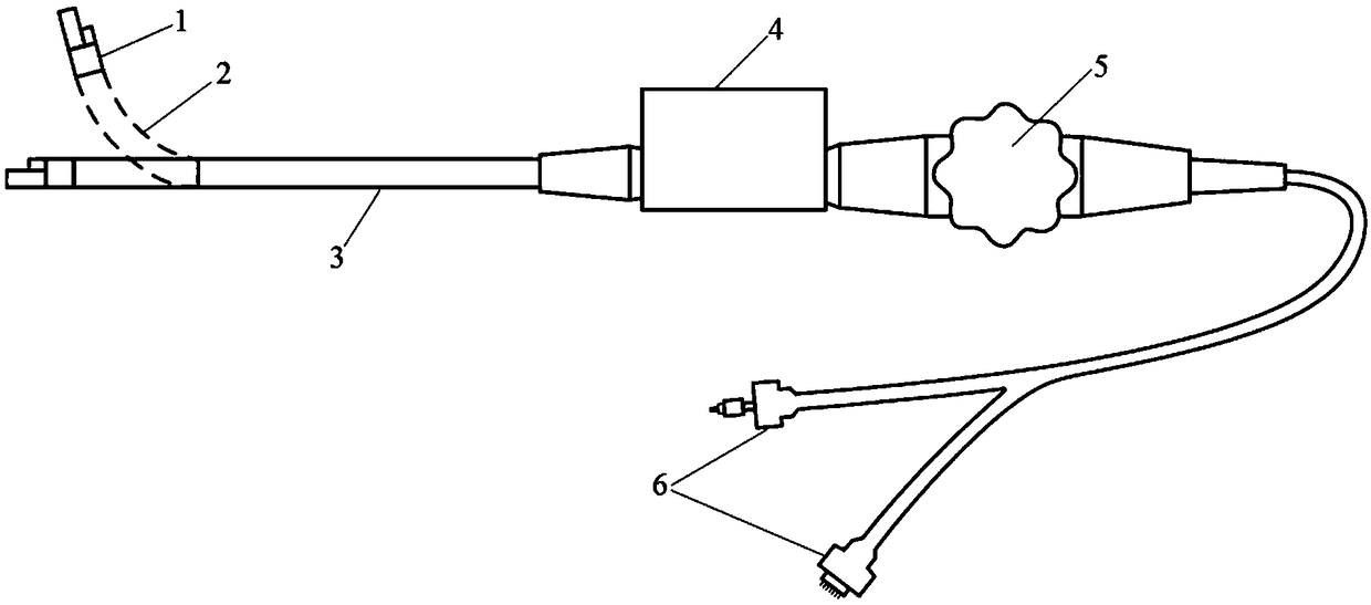
[0037] Such as figure 1 As shown, this embodiment is an endoscope for implementing a three-dimensional photoacoustic imaging method in a curved cavity based on snake bone direction change, including: an integrated scanning head 1, a snake bone bending part 2, an insertion hose 3, a three-dimensional Scanning part 4 , control handle 5 and joint part 6 .

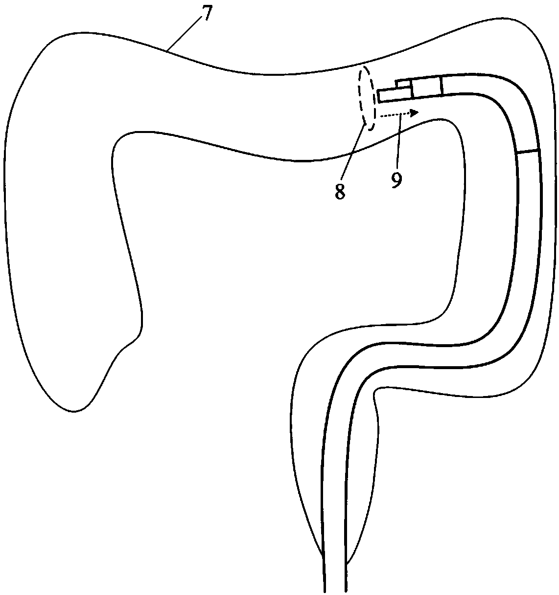
[0038] Such as figure 2 As shown, this embodiment is based on the three-dimensional photoacoustic imaging method in the curved cavity of the snake bone changing direction. By operating the control handle to adjust the four-way bending of the snake bone up, down, left, and right, the end of the endoscopic lens is bent accordingly. And under the guidance of the video image captured by the micro-optical camera, through the curved cavity 7, at the same time, the rotary motor and linear motor in the three-dimensional scanning part drive the photoacoustic probe to perform rotary scanning 8 and retraction scanning 9 to obtain the t...


PUM


Abstract

The invention discloses a snake-bone-turning-based bending cavity internal three-dimensional opto-acoustic endoscope and an imaging method thereof. The endoscope comprises an integral scanning head, asnake bone bending part, an insertion hose, a three-dimensional scanning part, a control handle and a joint part. An opto-acoustic scanning probe and an optical camera are integrated in the integralscanning head, the opto-acoustic probe achieves mechanical circular scanning and retracting through the three-dimensional scanning part, the snake bone bending part is connected with the integral scanning head and the insertion hose, under adjustment of the control handle, four-way bending of the integral scanning head is achieved, and the joint part is connected with a pulse laser and a main unit. According to the method, by means of snake bone turning, bending cavity internal three-dimensional opto-acoustic imaging under the guidance of video images is achieved. Accordingly, the snake-bone-turning-based bending cavity internal three-dimensional opto-acoustic imaging method and the endoscope thereof combine high-resolution opto-acoustic imaging with high-resolution optical imaging, and bymeans of snake bone turning, bending cavity internal surface high-definition video images and a three-dimensional structure, function and molecular images of cavity tissue are obtained.

Description

technical field [0001] The invention belongs to the technical field of medical endoscopes, and relates to a three-dimensional photoacoustic endoscope in a curved cavity based on snake bone direction change and an imaging method thereof. Background technique [0002] Medical endoscope is an instrument for inspection, diagnosis and treatment of human body cavity tissue. Existing endoscopes mainly include optical video endoscopes, ultrasonic endoscopes and optical coherence tomography endoscopes. Optical video endoscopy can provide high-definition imaging of the cavity tissue surface, but cannot observe the fault structure of cavity tissue, which limits its ability to diagnose diseases to a certain extent; ultrasonic endoscopy can obtain deep structural information of cavity tissue , but its imaging contrast is low and cannot reflect early lesions; optical coherence tomography endoscopy can image cavity sections through focused beams, and has high resolution, but its imaging d...

Claims


Application Information

Patent Timeline
Patent Type & Authority Applications(China)
IPC IPC(8): A61B1/005A61B1/04A61B1/06A61B1/07A61B8/12A61B5/00
CPCA61B1/0051A61B1/04A61B1/0684A61B1/07A61B5/0095A61B8/12
Inventor 杨思华金鑫熊科迪邢达
Owner SOUTH CHINA NORMAL UNIVERSITY
Why Eureka
Try Eureka