
Human respiration self-driven flexible respiration sensor and reparation method thereof

A breathing sensor, self-driven technology, applied in respirators, sensors, and evaluation of respiratory organs, etc., can solve problems such as expensive, harmful to the body, and narrow practical temperature range

Active Publication Date: 2019-01-08
UNIV OF ELECTRONICS SCI & TECH OF CHINA
View PDF7 Cites 13 Cited by
  • Summary
  • Abstract
  • Description
  • Claims
  • Application Information

AI Technical Summary

Problems solved by technology

[0006] The present invention aims at the problems of high price, narrow practical temperature range and the need to take oral medicine to harm the body in the traditional detection method. Self-Powered Breath Detection Sensor

Method used


Image

  • Human respiration self-driven flexible respiration sensor and reparation method thereof
  • Human respiration self-driven flexible respiration sensor and reparation method thereof
  • Human respiration self-driven flexible respiration sensor and reparation method thereof

Examples


Embodiment 1

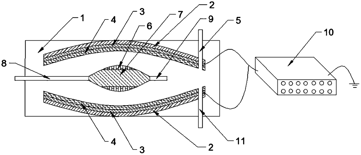
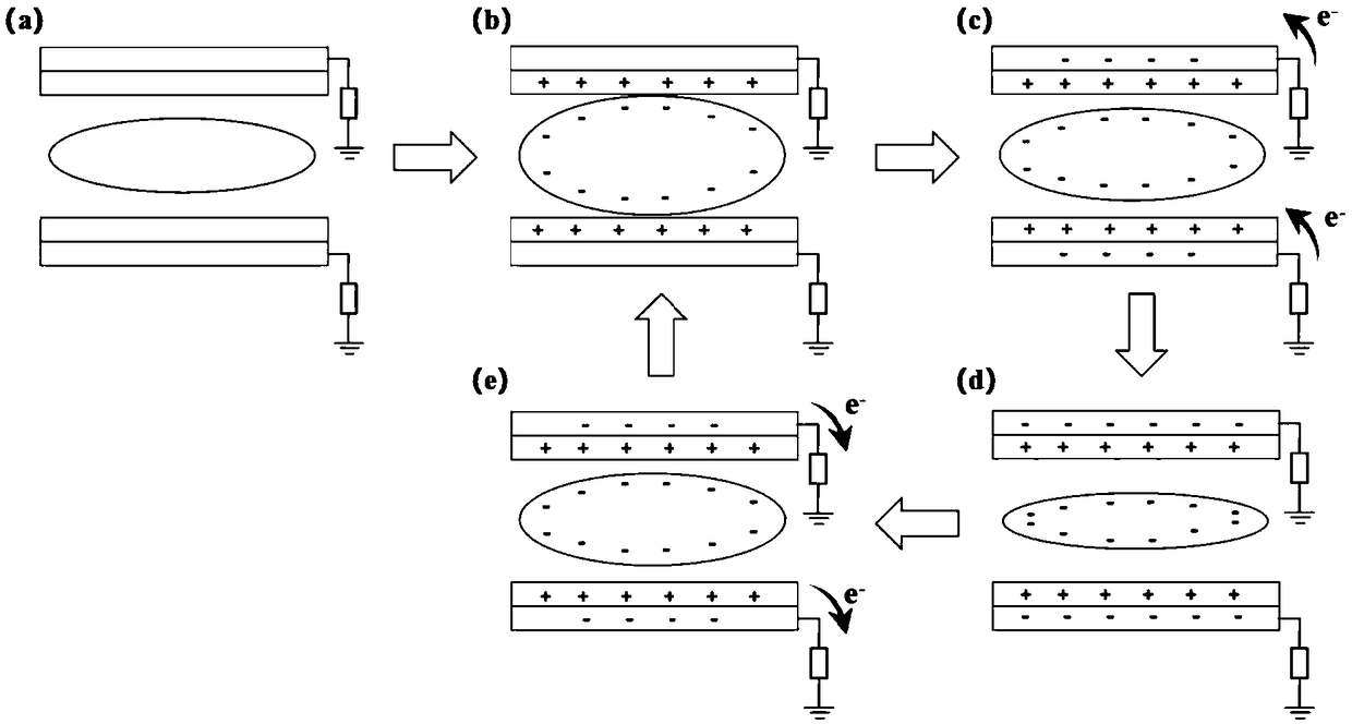
[0040] Such as figure 1 As shown, a flexible breathing sensor driven by human breathing includes a test chamber 1 and a digital electrometer 10. An upper detection component is arranged on the upper inner side wall of the test chamber 1, and a lower detection unit is arranged on the lower inner side wall of the test chamber 1. Components; the upper detection component and the lower detection component are symmetrically arranged up and down; the upper detection component includes a substrate 2, an electrode 3 and a first film 4 bonded sequentially from top to bottom, the first film 4 is a first friction film, and the substrate 2 Bonded with the upper inner side wall of the test chamber 1; a rubber airbag 7 is arranged between the upper detection assembly and the lower detection assembly, the rubber airbag 7 is bonded with a friction film 6, and the left end of the rubber airbag 7 is connected to a Intake cylinder 8, the right end of rubber airbag 7 is connected with outlet cyli...

Embodiment 2

[0055] Prepare a respiratory gas sensor for monitoring the target gas concentration in human breathing, and the detection steps are as follows:

[0056] (1): Take a 2mm thick polymethyl methacrylate plexiglass plate, cut it into a corresponding substrate by a laser cutting machine, and assemble it into a test cavity.

[0057] (2): Take a polyethylene terephthalate organic film with a thickness of 250 μm, wash it with chemical reagents such as acetone and ethanol, and dry it.

[0058] (3): Cut the cleaned polyethylene terephthalate organic film into a square substrate of 3 cm×3 cm by a laser cutting machine.

[0059] (4): A layer of gold electrode is vapor-deposited on the rectangular substrate by thermal evaporation to form an electrode, and the size of the electrode is 3cm×3cm.

[0060] (5): Assemble the polyaniline-metal oxide gas-sensing material by in-situ polymerization, and then attach a layer of polyaniline gas-sensing film on the surface of the gold electrode by air s...


PUM

PropertyMeasurementUnit
thicknessaaaaaaaaaa

Abstract

The invention discloses a human respiration self-driven flexible respiration sensor and belongs to the technical field of micro-electronic machinery. The sensor comprises a testing cavity and a digital electrometer, wherein an upper detection assembly and a lower detection assembly which are vertically symmetric are arranged on upper and lower inner side walls of the testing cavity respectively; the upper detection assembly comprises a base plate, an electrode and a first film which are bonded sequentially from top to bottom, and the base plate is bonded with the upper inner side wall of the testing cavity; a rubber gas bag is arranged in the testing cavity, a friction film is bonded on the rubber gas bag, the left end of the rubber gas bag is communicated with an gas inlet barrel, and theright end of the rubber gas bag is communicated with an gas outlet barrel; the symmetry axis of the rubber gas bag coincides with the vertical symmetry axis of the upper detection assembly and the lower detection assembly; the upper detection assembly and the lower detection assembly are both electrically connected with the digital electrometer which is grounded, an external power supply is not required, kinetic energy of respiratory gas is directly used to drive the rubber gas bag to expand, and the respiration frequency, respiration capacity and concentration of target gas are detected spontaneously.

Description

technical field [0001] The invention relates to the field of microelectronic machinery technology, in particular to a flexible breathing sensor driven by human breathing and a preparation method thereof. Background technique [0002] The composition of human respiratory gas is complex, except for N 2 , O 2 , CO 2 and H 2 In addition to these main components of O, there are hundreds of other trace gases. The change of breath composition comes from the metabolism of certain tissue structures inside the human body. Under normal circumstances, the concentration of these gases in the exhaled breath will remain within a certain range. However, if these tissue structures are lesions, there will inevitably be problems with their metabolism, causing some intermediate products to be excreted with the respiratory gas because they cannot be decomposed. Therefore, we can preliminarily diagnose diseases in the human body by detecting the concentration of the target gas in the exhaled...

Claims


Application Information

Patent Timeline
Patent Type & Authority Applications(China)
IPC IPC(8): G01N27/00A61B5/08A61B5/093
CPCA61B5/0816A61B5/093G01N27/00A61B5/083A61B5/087A61B5/082A61M16/024A61B5/113A61B5/4836A61M2016/0021
Inventor 太惠玲刘勃豪王斯袁震黄琦蒋亚东谢光忠
Owner UNIV OF ELECTRONICS SCI & TECH OF CHINA
Learn More