A retinal blood vessel morphology quantization method based on a connected region

A technology of retinal blood vessels and connected areas is applied in the field of retinal blood vessel morphology quantification based on connected areas to achieve the effect of improving simplicity and ensuring accuracy

Active Publication Date: 2019-01-08
CENT SOUTH UNIV
View PDF11 Cites 26 Cited by
  • Summary
  • Abstract
  • Description
  • Claims
  • Application Information

AI Technical Summary

Problems solved by technology

[0006] According to the topological structure of the retinal blood vessels, the present invention uses the intersection points of the retinal blood vessel network to traverse and determine the connected area of ​​the blood vessels, and measures the diameter and direction of the blood vessel segment on the basis of the connected domain of the blood vessels to solve the quantification problem of the retinal blood vessels

Method used


Image

  • A retinal blood vessel morphology quantization method based on a connected region
  • A retinal blood vessel morphology quantization method based on a connected region
  • A retinal blood vessel morphology quantization method based on a connected region

Examples


Embodiment Construction

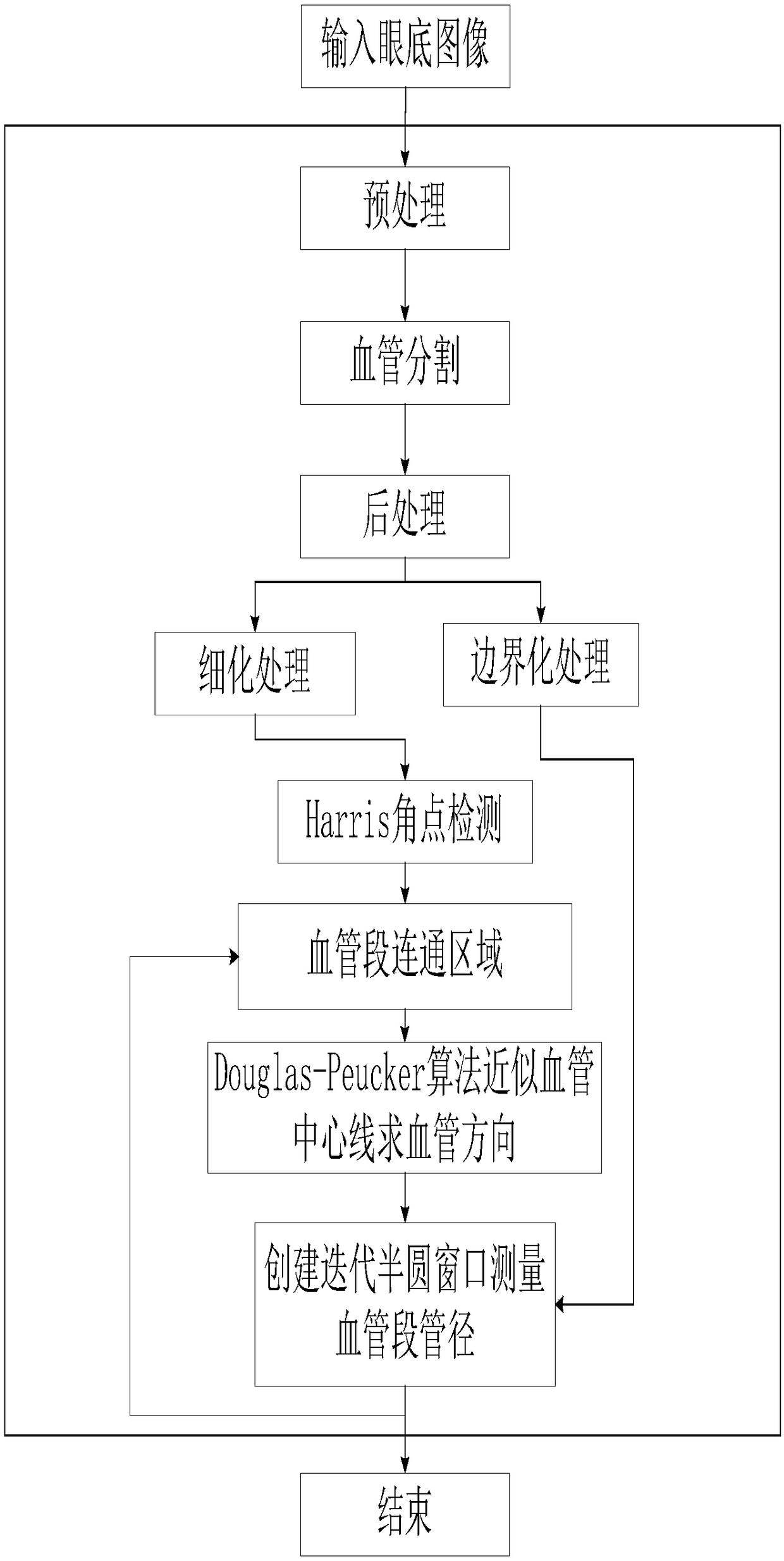
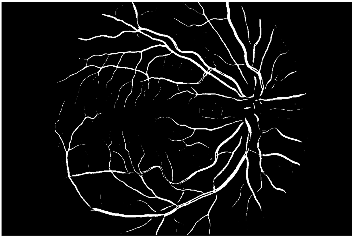
[0040] Such as figure 1 As shown, the embodiment of the present invention provides a method for quantifying retinal blood vessel morphology based on connected regions, including the following steps: the fundus image I src Carry out blood vessel segmentation processing, and use the method of fusion multi-label classification and deep learning to obtain retinal blood vessel segmentation map I seg ; Next, the blood vessel segmentation map I seg Perform post-processing to remove noise to obtain vascular network image I net ; and then respectively for I net Thinning and boundary operations are performed to obtain the corresponding vascular centerline network diagram I skl and Vessel Boundary Map I edge ; Then use the Harris corner detection algorithm to mark I skl vascular intersections and branch points in the I skl Remove the centerline map of the blood vessel segment I conn , and then the connected areas of blood vessel segments with different shapes and separated from ea...


PUM


Abstract

The invention provides a retinal blood vessel morphology quantization method based on a connected region. The method obtains a retinal blood vessel segmentation image after the fundus image is preprocessed, and then performs post-processing on the blood vessel segmentation image. On this basis, the vascular network is thinned and boundary treated, and the vascular centerline network and vascular boundary map are obtained. Corner detection is then performed and removed from the vascular centerline network so that the vascular segments of the vascular network form separate communication areas. Traversing is performed on the blood vessel segment, approximate the centerline of the blood vessel segments, and the blood vessel segment is changed into a broken line to calculate the direction of the blood vessel. At last, that initial diameter value is calculated, the center of the circle is selected by sliding on the centerline of the blood vessel segment, a semicircle window is created according to the direction of the circle cardiovascular and the diameter value measured in the early stage, and the distance between the window and the two intersection points of the blood vessel boundary is taken as a new diameter value. From this iteration, a set of vessel diameter values are measured, and the median value is the vessel diameter of the vessel segment. The invention is applicable to the quantification of large-scale retinal blood vessel morphology, and has high reliability.

Description

technical field [0001] The invention belongs to the technical field of image processing, and relates to a retinal blood vessel shape quantification method based on connected regions. technical background [0002] With the advancement of image capture hardware and the continuous development of computing efficiency, coupled with increasingly complex image analysis and machine learning techniques, it provides a basis for obtaining the tiny details of biological tissues in areas such as the retina, allowing professionals to detect images through image detection. An exception was found. The fundus retinal vascular image (hereinafter referred to as the fundus image) is the only image in the vascular system that can be directly obtained by non-invasive fundus photography, and the central retinal artery is the only small artery that can be directly observed in the living body. And related cardiovascular diseases will affect the retinal vascular structure, such as hypertension (incl...

Claims


Application Information

Patent Timeline
Patent Type & Authority Applications(China)
IPC IPC(8): G06T7/00G06T7/11G06N3/04
CPCG06T7/0012G06T7/11G06T2207/30101G06T2207/30041G06N3/045
Inventor 邹北骥戴玉兰朱承璋黄奕鑫胡蓉单希
Owner CENT SOUTH UNIV
Why Eureka
Try Eureka