
In vivo corneal parameter measuring device and measuring method based on optical coherence tomography

A technology of optical coherence tomography and measurement devices, which is applied in the field of measurement devices for body corneal parameters, can solve the problems of reducing the diagnostic sensitivity of keratoconus, not being able to distinguish the thickness of each layer of the cornea in detail, and being unable to be used as a reliable experimental method for corneal biomechanics, etc. To achieve the effect of overcoming inaccuracy, perfect function and accurate data

Pending Publication Date: 2019-01-11
WENZHOU MEDICAL UNIV
View PDF0 Cites 6 Cited by
  • Summary
  • Abstract
  • Description
  • Claims
  • Application Information

AI Technical Summary

Problems solved by technology

First, the parameters obtained by the instrument do not directly correspond to the standard constitutive parameters in physical mechanics, so they cannot be used as a reliable experimental method for measuring corneal biomechanical properties
Second, the instrument can only measure the biomechanical properties of the whole cornea, but cannot obtain the distribution map of the biomechanical properties of the cornea, thus reducing the sensitivity of keratoconus diagnosis
Third, the parameter measurement of the instrument is affected by the geometric parameters of the cornea (such as corneal thickness, curvature, etc.), which cannot truly reflect the biomechanical properties of the cornea
However, Pentacam cannot distinguish the thickness of each layer of the cornea in detail. For example, after LSAIK, corneal trauma, ulcer and keratoconus and other lesions, the thickness of the corneal flap and corneal stroma is particularly important in the diagnosis, treatment and evaluation of such diseases

Method used


Image

  • In vivo corneal parameter measuring device and measuring method based on optical coherence tomography
  • In vivo corneal parameter measuring device and measuring method based on optical coherence tomography
  • In vivo corneal parameter measuring device and measuring method based on optical coherence tomography

Examples


Embodiment Construction

[0058] In order to understand the technical content of the present invention more clearly, the following examples are given in detail.

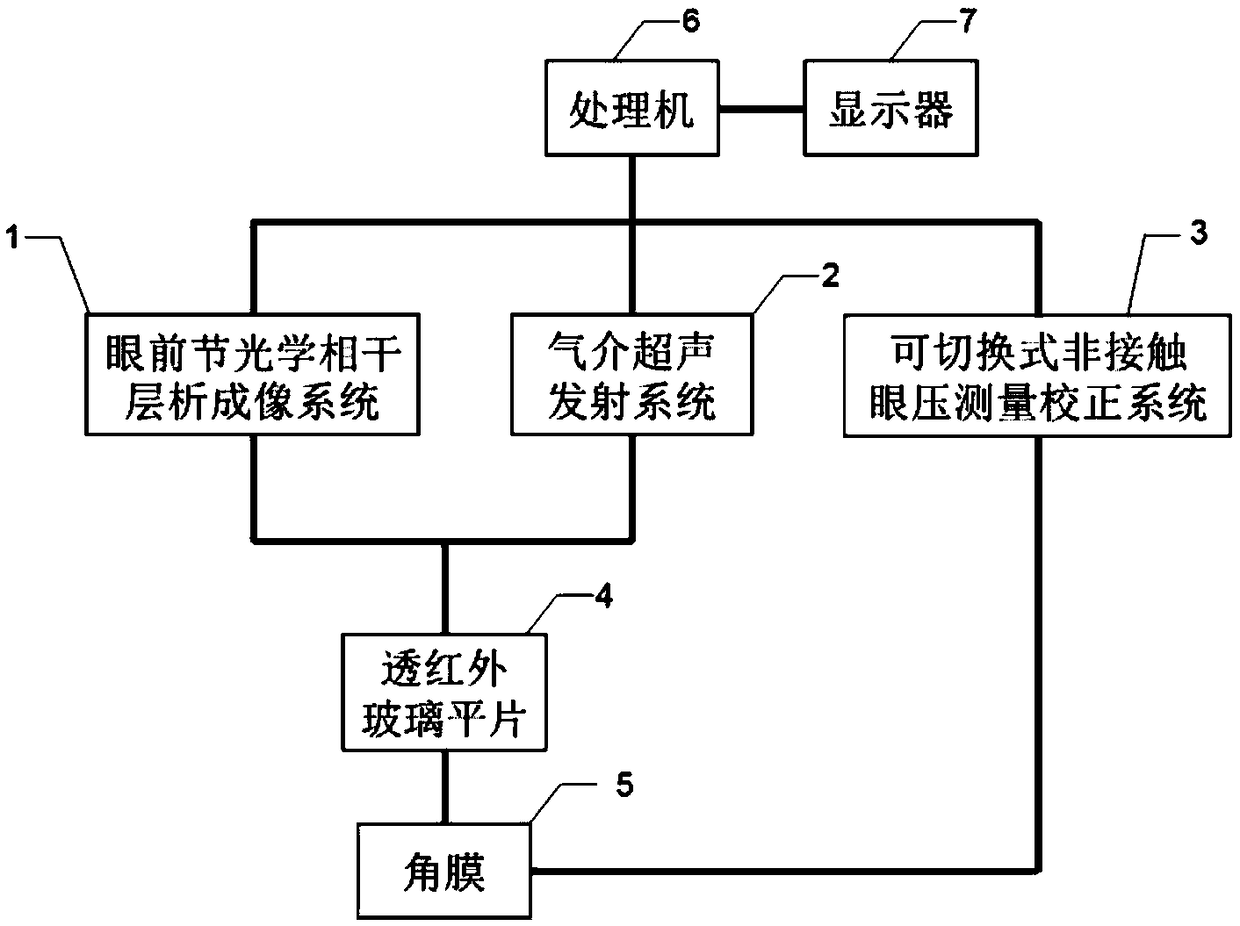
[0059] The device structure diagram of the present invention is as figure 1 As shown, it includes an anterior segment optical coherence tomography system 1 , an air-mediated ultrasound emission system 2 , a switchable non-contact intraocular pressure measurement correction system 3 , an infrared transparent glass flat film 4 , a cornea 5 , a processor 6 and a display 7 .

[0060] Among them, the anterior segment optical coherence tomography system 1 , the air-mediated ultrasound emission system 2 , and the switchable non-contact intraocular pressure measurement and correction system 3 are connected to the processor 6 respectively. The outgoing light beam of the anterior segment optical coherence tomography system 1 is transmitted through the infrared transparent glass flat sheet 4 to the cornea 5, and the light beam reflected from the cornea ...


PUM


Abstract

The invention relates to an in vivo corneal parameter measuring device and measuring method based on optical coherence tomography comprises an anterior segment optical coherence tomography system, anair-mediated ultrasonic emission system, a switchable non-contact intraocular pressure measuring correction system, an infrared transmissive glass plate, a processor, and a display. The corneal three-dimensional morphology images were obtained by the anterior segment optical coherence tomography system. When the ultrasound generated by the air-mediated ultrasound emission system caused the cornealresonance, the whole corneal elastic modulus was obtained from the corneal optical coherence tomography images loaded with the resonance ultrasound signal. At the same time, accord to the three-dimensional shape image of the cornea and the total corneal elastic modulus value, a switchable non-contact IOP measurement correction system is utilized to effectively correct the IOP value measure routinely to obtain an accurate IOP value, so as to overcome the inaccuracy of constant quantification processing of the corneal shape and biomechanical properties when the IOP is measured by the prior art.

Description

technical field [0001] The invention specifically relates to the technical field of measurement of in vivo corneal morphology, intraocular pressure and biomechanical performance parameters, in particular to a measurement device and method for in vivo corneal parameters based on optical coherence tomography technology. Background technique [0002] The measurement of intraocular pressure, corneal biomechanical properties and corneal morphology plays an important role in the diagnosis and treatment of glaucoma, corneal trauma, keratoconus and other ophthalmic diseases. However, under normal circumstances, the three are often closely related and influenced each other, so the integration of corneal morphology, biomechanical properties and intraocular pressure measurement is a very meaningful research content in the field of ophthalmic imaging examination. The integrated measurement method implemented in the present invention can provide more accurate treatment guidance and auxil...

Claims


Application Information

Patent Timeline
IPC IPC(8): A61B3/10A61B3/107A61B3/16
CPCA61B3/0008A61B3/102A61B3/107A61B3/165
Inventor 王媛媛沈梅晓朱德喜周煜恒吕帆
Owner WENZHOU MEDICAL UNIV
Learn More