Immunofluorescent kit for detecting different loci of HER2 antigen and application of immunofluorescent kit

A technology of immunofluorescence detection and detection process, which is applied in the field of immunofluorescence detection kits for different sites of human epidermal growth factor receptor 2 antigen, which can solve the problems of inability to perform dynamic detection and inability to judge the absence of extracellular segments

Inactive Publication Date: 2019-01-11
北京莱尔生物医药科技有限公司
View PDF3 Cites 7 Cited by
  • Summary
  • Abstract
  • Description
  • Claims
  • Application Information

AI Technical Summary

Problems solved by technology

This study detects the expression of full-length HER2 and the intracellular segment of HER2 in patient tissues, but cannot judge the absence of the extracellular segment, and needs to use two tissue sections respectively, so it can only be detected in patients whose tumor tissues can be observed and obtained, and cannot be detected. Perform dynamic detection

Method used


Image

  • Immunofluorescent kit for detecting different loci of HER2 antigen and application of immunofluorescent kit
  • Immunofluorescent kit for detecting different loci of HER2 antigen and application of immunofluorescent kit
  • Immunofluorescent kit for detecting different loci of HER2 antigen and application of immunofluorescent kit

Examples


Embodiment 1

[0079] Materials: smears of negatively enriched blood samples and SKBR3 smears for control cells.

[0080] Experimental steps:

[0081] 1. Draw 3.5ml of peripheral blood into an ACD (sodium citrate) anticoagulant tube. use The human peripheral blood leukocyte depletion kit negatively enriches tumor cells and fixes them on glass slides;

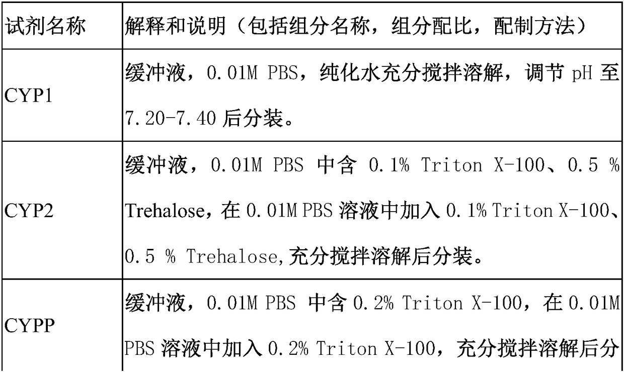
[0082] 2. Wash slides with CYP1 for 3 minutes x 3 times, 100-150 μL each time, to ensure that the entire sample area is covered;

[0083] 3. Absorb the excess liquid on the slide, add CYPP for 5 minutes, wash the slides with CYP1 as above for 3 minutes × 1 time; absorb excess liquid, add 200 μl of ice acetone:methanol (7:3) for 5 minutes, and wash the slides with CYP1 for 3 minutes × 3 times , to absorb excess water;

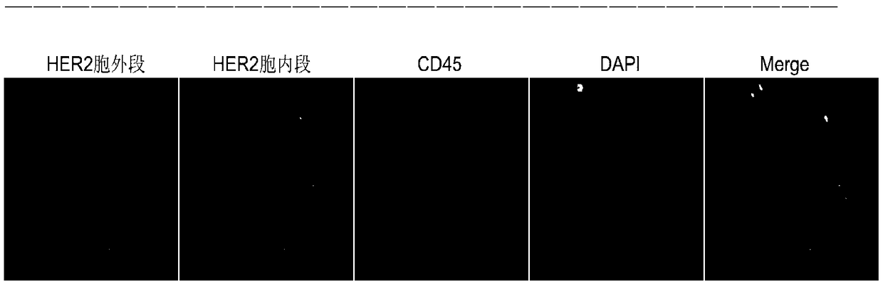
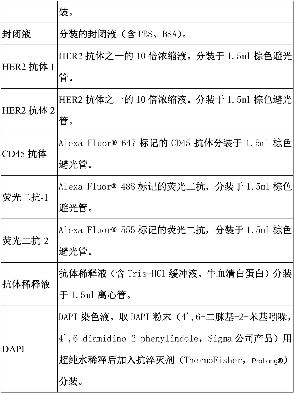
[0084] 4. Add 100-150 μl of blocking solution to block at room temperature for 25-30 minutes. Absorb excess blocking solution, add 100 μl of diluted HER2 antibody 1, HER2 antibody 2 and CD45 antibody, and incubate in a hum...

Embodiment 2

[0092] Materials: 1 tube of appropriate amount of anticoagulated blood, which is enriched by membrane filtration and then detected for protein. Experimental steps:

[0093] 1. Take an appropriate amount of peripheral blood and put it into a blood collection tube containing anticoagulant, and shake it slightly to mix.

[0094] 2. Add the suspension to the membrane filtration separation tumor cell technology device, and slowly pass through the filter and the filter membrane.

[0095] 3. After the filtration is completed, continue to add 50ml of 0.01M PBS to the membrane filtration device, wash the cell suspension attached around the tube wall into the membrane filtration device, and let it pass through the filter and membrane;

[0096] 4. Fix the cells on the filter membrane;

[0097] 5. Perform the same operation as in Example 1 to detect the protein.

Embodiment 3

[0099] Materials: 1 tube of appropriate amount of anticoagulated blood, which is enriched by microfluidic method and then detected for protein. Experimental steps:

[0100] 1. The appropriate amount of blood drawn is enriched using microfluidic chips of various principles.

[0101] 2. After enrichment, the samples were subjected to protein immunofluorescence detection.


PUM


Abstract

The invention relates to an immunofluorescent kit for detecting different loci of an HER2 antigen and an application of the immunofluorescent kit. A detection principle of the method disclosed by theinvention is as follows: firstly, enriching non-humoral rare nucleated cells, and then, detecting the expression of target proteins in the enriched cells in cells by using an immunofluorescene detection method according to an antigen-antibody reaction principle. According to the kit, a common antigen CD45 of a target cell and a leukocyte is fluorescently labeled, and the cells with positive targetproteins and negative CD45 are screened, so that the non-humoral rare nucleated cells with positive specific proteins in the blood are interpreted and counted.

Description

Technical field: [0001] The invention relates to a medical detection method, in particular to the detection of tumor cells in body fluids, in particular to an immunofluorescence detection kit for different sites of human epidermal growth factor receptor 2 (HER2) antigen, which is suitable for detection of HER2 in different types of samples, and also It relates to the application of the kit in detecting different sites of HER2 antigen. Background technique: [0002] At present, the incidence of cancer is relatively high, which seriously threatens human health. Some cancers are difficult to detect early and are often diagnosed at an advanced stage, such as lung cancer, colorectal cancer, ovarian cancer, and pancreatic cancer. Correspondingly, the 5-year survival rate of advanced cancer is significantly lower than that of early-stage cancer, and early detection, early diagnosis and precise treatment can significantly prolong the survival time of patients. [0003] Human epide...

Claims


Application Information

Patent Timeline
Patent Type & Authority Applications(China)
IPC IPC(8): G01N33/574G01N33/68G01N33/533G01N21/64
CPCG01N21/6428G01N33/533G01N33/57496G01N33/6803G01N2800/7028
Inventor 郭素杰郭志敏樊晓婷
Owner 北京莱尔生物医药科技有限公司
Why Eureka
Try Eureka