Magnetic resonance imaging method and system

A magnetic resonance imaging and imaging sequence technology, which is applied in the direction of using nuclear magnetic resonance imaging system for measurement, magnetic resonance measurement, and magnetic variable measurement, can solve the problems of long time consumption of cardiac images and low imaging efficiency, and reduce data acquisition. Time-consuming, improving imaging efficiency, and reducing the number of acquisitions

Active Publication Date: 2019-03-22
SHANGHAI UNITED IMAGING HEALTHCARE
View PDF8 Cites 10 Cited by
  • Summary
  • Abstract
  • Description
  • Claims
  • Application Information

AI Technical Summary

Problems solved by technology

[0005] In short, it takes a long time to acquire images of the whole heart using existing MRI techniques, resulting in low imaging efficiency

Method used


Image

  • Magnetic resonance imaging method and system
  • Magnetic resonance imaging method and system
  • Magnetic resonance imaging method and system

Examples


Embodiment 1

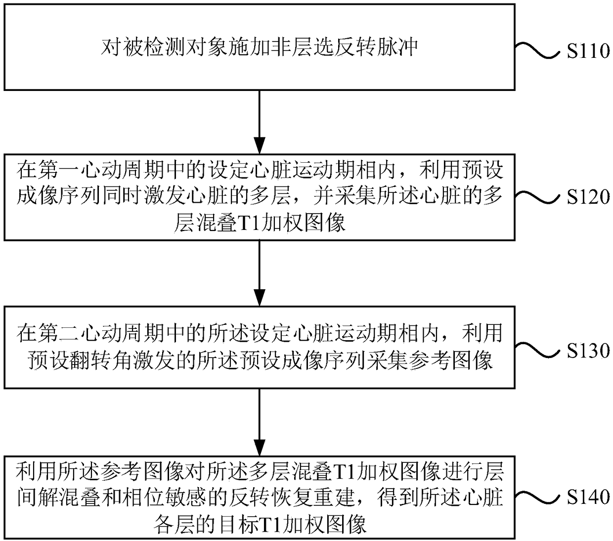
[0029] The magnetic resonance imaging method provided in this embodiment is applicable to fast magnetic resonance imaging of the heart. The method can be performed by a magnetic resonance imaging device, the device can be realized by software and / or hardware, and the device can be integrated in a medical device that can perform magnetic resonance imaging. see figure 1 , the method of this embodiment specifically includes the following steps:

[0030] S110, applying a non-layer selection inversion pulse to the detected object.

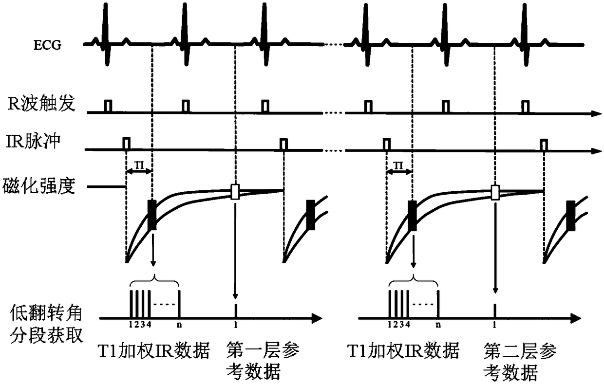
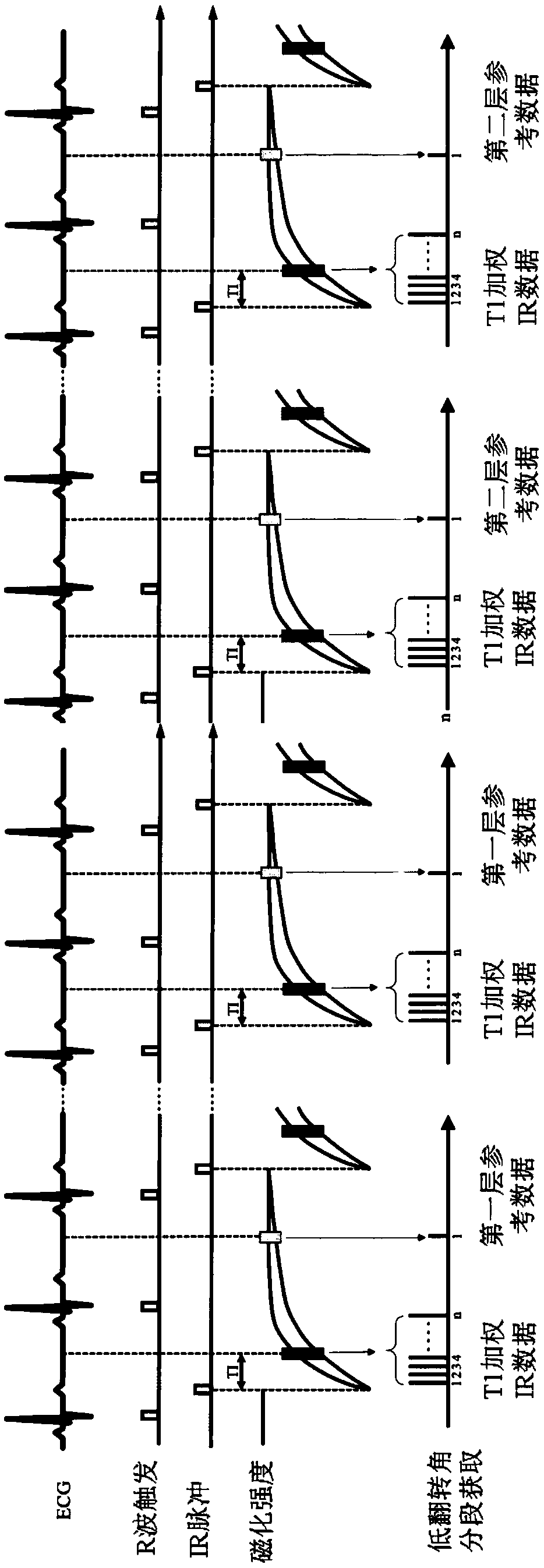
[0031] Specifically, in the process of magnetic resonance imaging of the heart, in order to obtain a stable and effective heart image, the automaticity of the heart movement is used to select the timing of data acquisition. During specific implementation, the extraction is in the static magnetic field B 0 The segmented electrocardiograph (ECG) of the detected object is used as a trigger signal, and a non-layer-selected inversion pulse is applied to t...

Embodiment 2

[0050] In this embodiment, on the basis of the first embodiment above, "use the reference image to perform interlayer de-aliasing and phase-sensitive inversion recovery reconstruction on the multi-layer aliased T1-weighted image to obtain the The target T1-weighted image" was further optimized. The explanations of terms that are the same as or corresponding to the above-mentioned embodiments will not be repeated here. see image 3 , the magnetic resonance imaging method provided in this embodiment includes:

[0051] S310, applying a non-layer selection inversion pulse to the detected object.

[0052] S320. In a set cardiac motion phase in the first cardiac cycle, use a preset imaging sequence to simultaneously excite multiple layers of the heart, and acquire an aliased T1-weighted image of the multiple layers of the heart.

[0053] S330. In the set cardiac motion phase in the second cardiac cycle, use the preset imaging sequence excited by a preset flip angle to acquire a r...

Embodiment 3

[0068] In this embodiment, on the basis of the foregoing embodiments, the "multi-layer aliased T1-weighted image" is further limited. On this basis, further carry out "using the reference image to perform interlayer de-aliasing and phase-sensitive inversion recovery reconstruction on the multi-layer aliased T1-weighted image to obtain the target T1-weighted image of each layer of the heart" Optimized. The explanations of terms that are the same as or corresponding to the above-mentioned embodiments will not be repeated here. see Figure 4 , the magnetic resonance imaging method provided in this embodiment includes:

[0069] S410, applying a non-layer selection inversion pulse to the detected object.

[0070] S420. In the set cardiac motion phase in the first cardiac cycle, use a preset imaging sequence to simultaneously excite multiple layers of the heart, and acquire a multi-layer aliased T1-weighted image of the heart, the multi-layer aliased T1 The K-space corresponding...


PUM


Abstract

Embodiments of the invention disclose a magnetic resonance imaging method and system. The method comprises the following steps: applying non-layering inverse pulses to a detected object; simultaneously stimulating multiple layers of a heart by using a preset imaging sequence in a set heart movement phase in a first heartbeat period, and acquiring a multilayer aliasing T1 weighted image of the heart; acquiring a reference image by using a preset imaging sequence stimulated by a preset overturning angle in a set heart movement phase in a second heartbeat period, wherein the first heartbeat period and the second heartbeat period correspond to a same breath-holding and are two adjacent heartbeat periods, and the reference image corresponds to one layer of the multiple layers; and performing the interlayer de-aliasing and phase-sensitive reverse restoration re-establishment for the multilayer aliasing T1 weighted image by using the reference image to obtain a target T1 weighted image of each layer of the heart. By virtue of the technical scheme, on the premise of ensuring the image quality, the magnetic resonance data acquisition efficiency can be improved.

Description

technical field [0001] Embodiments of the present invention relate to medical magnetic resonance technology, and in particular to a magnetic resonance imaging method and system. Background technique [0002] Myocardial infarction (abbreviated as myocardial infarction) is a serious heart disease. After myocardial infarction, for the myocardium whose myocardial activity has not completely disappeared (called salvageable myocardium), cardiac function can be improved by restoring myocardial perfusion, while myocardial perfusion of infarcted myocardium without myocardial activity will induce fatal heart rhythm out of order. Therefore, it is necessary to assess myocardial viability to distinguish salvageable myocardium from infarcted myocardium, so as to assess the probability of myocardial function recovery after reperfusion. [0003] At present, magnetic resonance imaging (MRI) is considered to be one of the effective means to evaluate myocardial activity. To better assess my...

Claims


Application Information

Patent Timeline
Patent Type & Authority Applications(China)
IPC IPC(8): G01R33/48A61B5/055
CPCA61B5/055G01R33/48
Inventor 徐健张哲豪谭国陞娄昕马林
Owner SHANGHAI UNITED IMAGING HEALTHCARE
Why Eureka
Try Eureka