
Choroidal angiography method and device based on optical coherence tomography imaging body scanning

A technology of optical coherence tomography and angiography, which is applied in the field of biomedical image processing, can solve the problems of unfavorable image processing segmentation and extraction, poor morphology of choroidal blood vessels, and influence on extraction and analysis of choroidal blood vessels, so as to facilitate extraction and quantitative research , The effect of eliminating the shadow of retinal blood vessels

Inactive Publication Date: 2019-05-10
CIXI INST OF BIOMEDICAL ENG NINGBO INST OF MATERIALS TECH & ENG CHINESE ACAD OF SCI +1
View PDF7 Cites 7 Cited by
  • Summary
  • Abstract
  • Description
  • Claims
  • Application Information

AI Technical Summary

Problems solved by technology

[0005] 1. The detection sensitivity of OCT decreases with the detection depth and the index of detection signal transmission in the tissue;
[0006] 2. Retinal vascular shadow affects the extraction and analysis of choroidal vessels;
[0007] 3. Choroidal blood flow velocity is much greater than retinal blood flow velocity, which has adverse effects on OCT and OCT angiography;
[0008] 4. The morphology of choroidal blood vessels is not as clear as that of retinal blood vessels, which is not conducive to segmentation and extraction in image processing

Method used


Image

  • Choroidal angiography method and device based on optical coherence tomography imaging body scanning
  • Choroidal angiography method and device based on optical coherence tomography imaging body scanning
  • Choroidal angiography method and device based on optical coherence tomography imaging body scanning

Examples


Embodiment 1

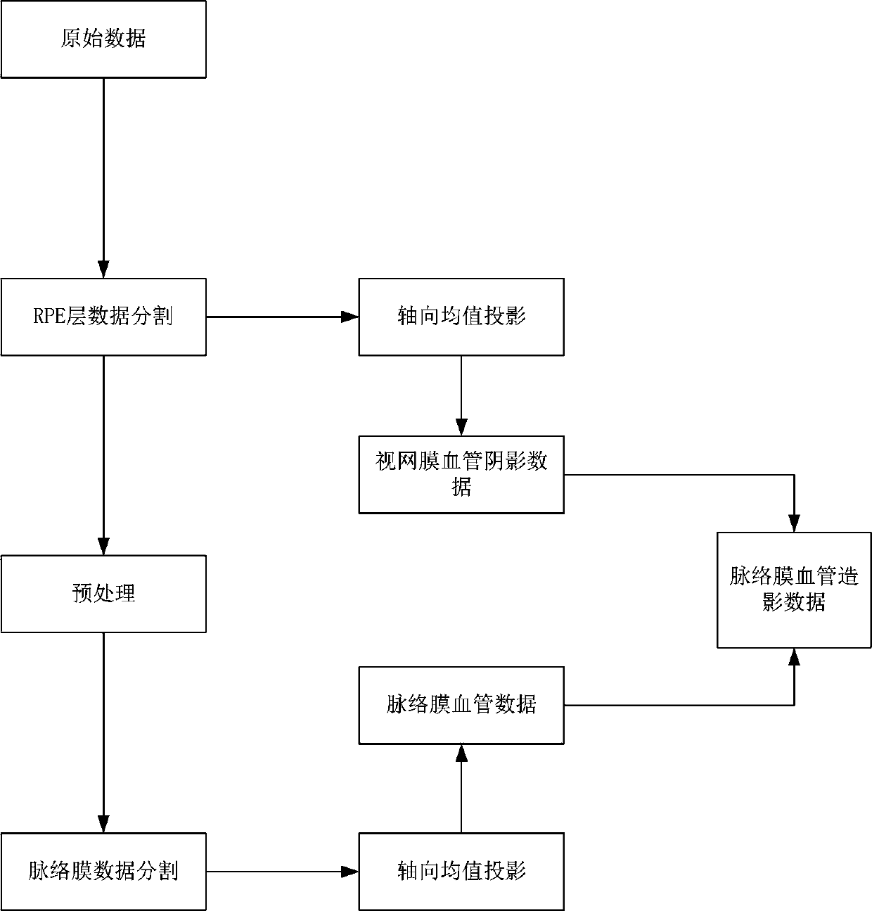
[0044] A choroidal angiography method based on optical coherence tomography volume scanning, its implementation steps can be divided into:

[0045] Step S1, obtaining the original data of the OCT volume scan of the fundus. Raw data are acquired using optical coherence tomography (OCT) equipment that examines samples. Embodiments described in this disclosure can use any OCT device to acquire raw data.

[0046] Step S2, extracting retinal vessel shadow data and choroidal vessel data from the original data. Among them, the extraction of retinal blood vessel shadow data mask includes:

[0047] Step B-1, segmenting the retinal pigment epithelium (RPE) layer data from the original data. The segmentation method of retinal pigment epithelium data can be a method based on threshold, a method based on graph theory and a method based on machine learning. In this embodiment, a method based on graph theory is used to segment the original data to obtain RPE data.

[0048] Step B-2, per...

Embodiment 2

[0066] A choroidal angiography device based on optical correlation tomography volume scanning, which implements the radiography method described in one embodiment to perform choroidal angiography.


PUM


Abstract

The invention relates to the field of biomedical image processing, in particular to a choroidal angiography method based on optical coherence tomography imaging body scanning. The method can be widelyused for screening and diagnosis of medical ophthalmic diseases. The method includes the steps of S1, acquiring original data of OCT body scanning fundus; S2, extracting retinal blood vessel shadow data and choroidal blood vessel data from the original data; S3, using the retinal blood vessel shadow data for masking the choroidal blood vessel data to obtain choroidal angiography data. According to the technical scheme above, by extracting the retinal blood vessel shadow data to mask the choroidal blood vessel data, retinal blood vessel shadows in the choroidal blood vessel data are removed, so that the influence of the retinal blood vessel shadows on the contrast and continuity of choroidal blood vessel projection images is reduced, and choroidal vessels can be completely and clearly displayed.

Description

technical field [0001] The invention relates to the field of biomedical image processing, in particular to a choroidal angiography method based on optical coherence tomography volume scanning, which can be widely used in the screening and diagnosis of medical ophthalmic diseases. Background technique [0002] The choroid plays a very important role in the blood oxygen supply and metabolism of the human eye. In addition to pathological changes in the choroid such as choroidal neovascularization, eye diseases such as pathological myopia, glaucoma, diabetic retinopathy, etc., and brain diseases such as Alzheimer's disease, stroke, etc. are closely related to many characteristics of the choroid, such as choroid Layer thickness, vascular distribution, etc. [0003] Optical coherence tomography (OCT) is a non-contact three-dimensional fundus imaging modality, which has been widely used in the screening, diagnosis and treatment of ophthalmic diseases. Unlike multispectral fundus ...

Claims


Application Information

Patent Timeline
IPC IPC(8): A61B3/12A61B3/14A61B5/00
Inventor 杨建龙毛浩宇蒋珊珊赵一天程骏胡衍刘江
Owner CIXI INST OF BIOMEDICAL ENG NINGBO INST OF MATERIALS TECH & ENG CHINESE ACAD OF SCI
Learn More