Magnetic immunochemiluminescence detection method for PD-L1 exosomes

A chemiluminescence detection, PD-L1 technology, applied in chemiluminescence/bioluminescence, analysis by chemical reaction of materials, biological testing, etc., can solve the problem of low specificity of exosome detection and low efficiency of magnetic immune separation , inconvenient operation and other problems, to achieve the effect of increasing the incubation and mixing effect, fast detection, and convenient operation

Inactive Publication Date: 2019-05-21
HUAZHONG UNIV OF SCI & TECH
View PDF3 Cites 4 Cited by
  • Summary
  • Abstract
  • Description
  • Claims
  • Application Information

AI Technical Summary

Problems solved by technology

[0005] The invention solves the technical problems of low exosome detection specificity, inconvenient operation and high cost in the prior art, as well as the technical problem of low efficiency of magnetic immune separation

Method used


Image

  • Magnetic immunochemiluminescence detection method for PD-L1 exosomes
  • Magnetic immunochemiluminescence detection method for PD-L1 exosomes

Examples


Embodiment 1

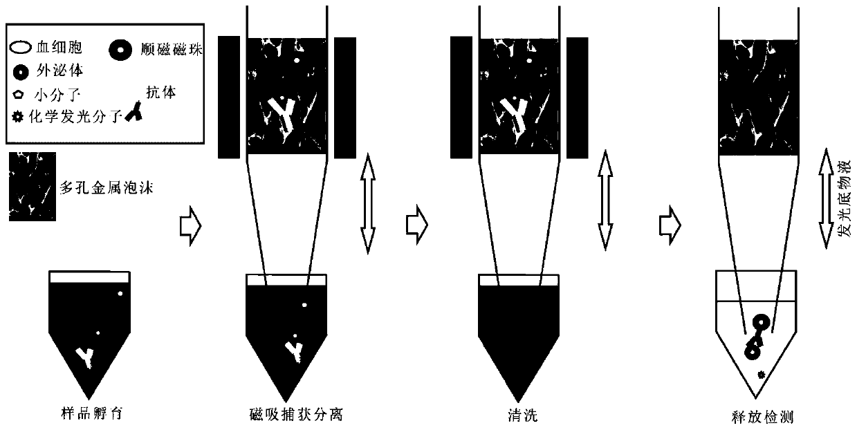
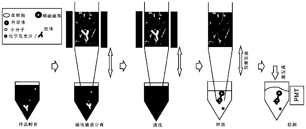
[0031] A magnetic immunochemiluminescent detection kit for PD-L1 exosomes, comprising exosome antibodies labeled with chemiluminescence molecules, exosome antibodies labeled with paramagnetic beads, hollow columns filled with porous metal foam, and magnets; The antibody on the chemiluminescent molecule-labeled antibody is an exosome PD-L1 antibody, and the antibody on the magnetic bead-labeled antibody is an exosome PD-L1 antibody. The porous metal foam is porous iron foam. The diameter of the pores in the porous iron foam is 10um. The chemiluminescent molecule is horseradish peroxidase. The diameter of the paramagnetic beads is 10 nm.

Embodiment 2

[0033] A magnetic immunochemiluminescent detection kit for PD-L1 exosomes, comprising exosome antibodies labeled with chemiluminescence molecules, exosome antibodies labeled with paramagnetic beads, hollow columns filled with porous metal foam, and magnets; The antibody on the chemiluminescent molecule-labeled antibody is an exosome PD-L1 antibody, and the antibody on the magnetic bead-labeled antibody is an exosome CD63 antibody. The porous metal foam is porous nickel foam. The diameter of the pores in the porous nickel foam is 50um. The chemiluminescent molecule is acridine ester. The diameter of the paramagnetic beads is 50nm.

Embodiment 3

[0035] A magnetic immunochemiluminescent detection kit for PD-L1 exosomes, comprising exosome antibodies labeled with chemiluminescence molecules, exosome antibodies labeled with paramagnetic beads, hollow columns filled with porous metal foam, and magnets; The antibody on the chemiluminescent molecule-labeled antibody is an exosome CD63 antibody, and the antibody on the magnetic bead-labeled antibody is an exosome PD-L1 antibody. The porous metal foam is porous cobalt foam. The diameter of the pores in the porous cobalt foam is 100um. The chemiluminescent molecule is horseradish peroxidase. The diameter of the paramagnetic beads is 1000 nm.


PUM

PropertyMeasurementUnit
diameteraaaaaaaaaa
diameteraaaaaaaaaa
diameteraaaaaaaaaa

Abstract

The invention discloses a magnetic immunochemiluminescence detection method for PD-L1 exosomes and belongs to the technical field of rapid in-vitro detection. A kit used in detection contains an antibody A, an antibody B, a porous metal foam-filled hollow column, and a magnet; the antibody A is a chemiluminescent molecule exosomal antibody or an enzyme-labeled exosomal antibody which is capable ofmaking a substrate emit light; the antibody B is a magnetic bead-labeled exosomal antibody; at least one of the antibody A and antibody B is an exosome PD-L1 antibody. The detection method includes the following steps that: the antibody A, the antibody B and a sample are incubated; efficient capture and separation are realized by means of the porous metal foam magnet; and after impurities are cleaned, a magnetic field is removed, and a double-antibody sandwiched composite is collected and detected. The method integrates sample incubation, magnet efficient capture and separation, and rapid detection, and has the advantages of convenient operation, rapid detection, low cost and the like.

Description

technical field [0001] The invention belongs to the technical field of in vitro rapid detection, and in particular relates to a magnetic immunochemiluminescent detection kit and detection method for PD-L1 exosomes. Background technique [0002] Exosomes are nanoscale vesicles (50-150nm in diameter) secreted by cells into the extracellular microenvironment, which were first discovered in 1983. However, due to the lack of analytical means, exosomes have long been considered as a tool for cells to excrete intracellular garbage. With the continuous innovation of analytical methods, the mechanism of exosome secretion has been gradually revealed. Now, the mainstream view in the scientific community is that exosomes are mainly involved in intercellular signal transmission and as disease markers. The 2013 Nobel Prize in Physiology or Medicine was awarded to three outstanding scientists who revealed major discoveries in this field. However, the resulting exosome function research ...

Claims


Application Information

Patent Timeline
Patent Type & Authority Applications(China)
IPC IPC(8): G01N33/68G01N21/76
Inventor 刘笔锋陈鹏
Owner HUAZHONG UNIV OF SCI & TECH
Why Eureka
Try Eureka