A three-dimensional spherical index determination method based on a CTA image

A technology of sphericity index and measurement method, which is applied in the field of medical image processing, can solve problems such as low quality of cardiac ultrasound images, unstable analysis results, and unfavorable observation of diseases, and achieves easy control, low degree of manual participation, and easy clinical use Effect

Active Publication Date: 2019-05-21
XIDIAN UNIV
View PDF20 Cites 2 Cited by
  • Summary
  • Abstract
  • Description
  • Claims
  • Application Information

AI Technical Summary

Problems solved by technology

2D ultrasound imaging is planar imaging and cannot truly reflect the three-dimensional changes in the shape of the left ventricle
However, to date, there is no method for assessing cardiac morphological structural changes using cardiac CTA
[0003] To sum up, the problems existing in the existing technology are: the current analysis of cardiac CTA images is mostly limited to coronary a

Method used


Image

  • A three-dimensional spherical index determination method based on a CTA image
  • A three-dimensional spherical index determination method based on a CTA image
  • A three-dimensional spherical index determination method based on a CTA image

Examples


Embodiment Construction

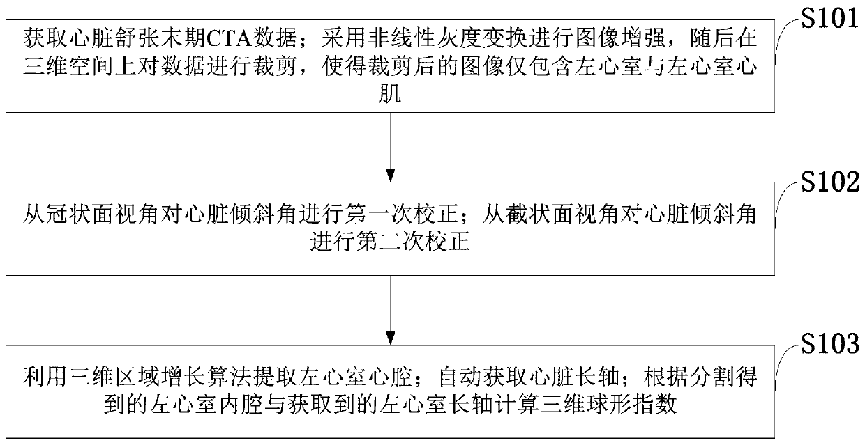
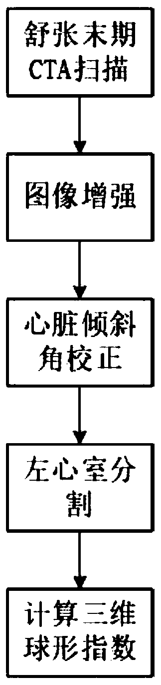
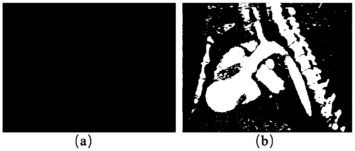
[0042] In order to make the object, technical solution and advantages of the present invention more clear, the present invention will be further described in detail below in conjunction with the examples. It should be understood that the specific embodiments described here are only used to explain the present invention, not to limit the present invention.

[0043]The current analysis of cardiac CTA images is mostly limited to coronary artery lesions, while ignoring the potentially usable and discoverable information of cardiac CTA images. Cardiac ultrasound, as a commonly used medical image for clinical analysis of cardiac morphology, has problems such as low image quality and unstable analysis results; there is no method for evaluating cardiac morphology and structural changes using cardiac CTA. For the first time, the present invention uses end-diastolic CTA images to evaluate changes in cardiac morphology and structure. The difficulty lies in how to minimize human involveme...


PUM


Abstract

The invention belongs to the technical field of medical image processing, and discloses a three-dimensional spherical index determination method based on a CTA image. The method includes: Obtaining CTA data at the final diastole stage; Carrying out image enhancement by adopting nonlinear gray scale transformation, and then clipping data in a three-dimensional space, so that the clipped image onlycomprises the left ventricle and the left ventricle myocardium; Correcting the heart inclination angle for the first time from the coronal plane view angle; Correcting the heart inclination angle forthe second time from the perspective of the section surface; Extracting a left ventricle heart cavity by using a three-dimensional region growing algorithm; Automatically obtaining a heart long axis;And calculating a three-dimensional spherical index according to the left ventricle inner cavity obtained through segmentation and the obtained left ventricle long axis. According to the method, an automatic measurement method is provided for evaluation of centrifugal reconstruction caused by overload or integral myocardial reconstruction caused by non-acute myocardial infarction, and an accurateand robust ventricular shape change degree quantification result is provided.

Description

technical field [0001] The invention belongs to the technical field of medical image processing, and in particular relates to a method for measuring a three-dimensional spherical index based on a CTA (CT angiography) image. Background technique [0002] At present, the existing technology commonly used in the industry is as follows: after myocardial infarction, part of the myocardial ischemia necrosis, loss of systolic function, uncoordinated contraction and movement of the left ventricular wall, resulting in the ineffective emptying of the blood in the left ventricular cavity, and the increase of residual blood , the pressure load in the ventricle increases, and the stress on the ventricular wall during systole and diastole increases, causing the expansion of the entire left ventricle (including the infarcted area and non-infarcted area) and changes in the structure and shape of the ventricle, which is left ventricular remodeling. Left ventricular remodeling is a precursor ...

Claims


Application Information

Patent Timeline
IPC IPC(8): G06T7/00G06T7/11G06T7/13G06T7/136G06T7/187G06T7/60G06T5/00
Inventor 梁继民徐真真陶博曹丰任胜寒陈雪利胡海虹
Owner XIDIAN UNIV
Why Eureka
Try Eureka