Ultrasonic image processing method and system, equipment and storage medium

An ultrasound image and processing method technology, applied in the field of ultrasound, can solve the problems of high daily working hours, poor consistency of inspection results, difficulty in satisfying mammary gland inspection, etc., to assist body tissue inspection, relieve doctor vacancies, and improve detection efficiency and the effect on accuracy

Pending Publication Date: 2019-05-31
清影医疗科技(深圳)有限公司
View PDF6 Cites 5 Cited by
  • Summary
  • Abstract
  • Description
  • Claims
  • Application Information

AI Technical Summary

Problems solved by technology

Ultrasound examination is a detection method suitable for Chinese women's breast characteristics. The existing detection method relies on doctors to perform breast ultrasound examination, but the accuracy rate of reading films with naked eyes is only 60-70%, and the reading time for each film is about 10 minutes. In addition, due to the influence of factors such as doctor's experience and working status, the consistency of traditional film reading with naked eyes is poor; moreover, traditional film reading is limited by the number of doctors and daily working hours, and it is difficult to meet the requirements of breast examination. need

Method used


Image

  • Ultrasonic image processing method and system, equipment and storage medium
  • Ultrasonic image processing method and system, equipment and storage medium
  • Ultrasonic image processing method and system, equipment and storage medium

Examples


Embodiment 1

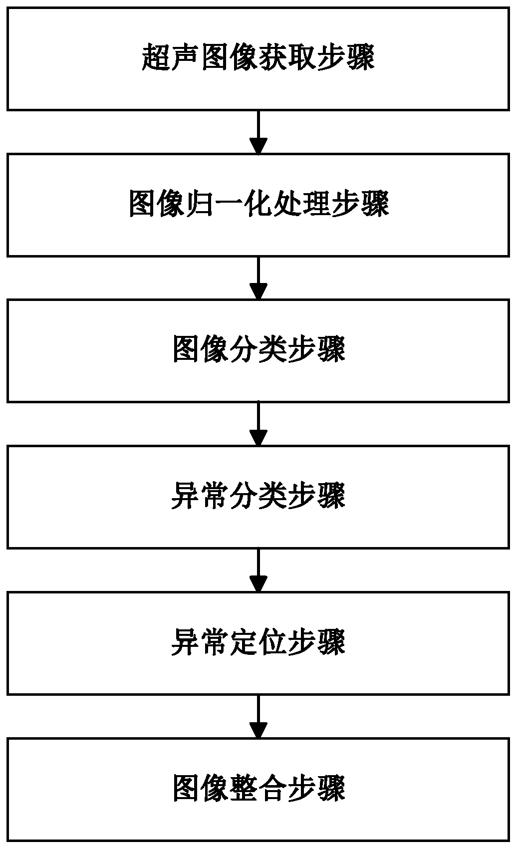
[0037] A kind of ultrasonic image processing method, reference figure 1 , figure 1 It is a flow chart of a specific embodiment of an ultrasonic image processing method in the present invention, comprising the following steps:

[0038] (1) Ultrasound image acquisition step: acquire multiple ultrasound images according to the ultrasound video of the body tissue under test, and extract multiple frames of images from the dynamic ultrasound video as the ultrasound image, wherein the body tissue under test includes breast and thyroid gland;

[0039] (2) Image normalization processing step: Normalize the format of the ultrasound image. Because the parameters of the ultrasound equipment are different, the quality of the ultrasound video obtained is also different. Therefore, setting this step can solve the problem of images of different ultrasound equipment. Differences lead to inaccurate processing results and improve the accuracy of subsequent processing results.

[0040] (3) Imag...

Embodiment 2

[0047] An ultrasonic image processing system, comprising:

[0048] An ultrasound image acquisition unit, configured to perform the ultrasound image acquisition step, and acquire multiple ultrasound images according to the ultrasound video of the body tissue under test, where the body tissue under test includes breast and thyroid gland;

[0049] The image normalization processing unit is used to perform the image normalization processing step, and normalize the format of the ultrasonic image;

[0050] The image classification unit is used to perform the image classification step, and obtain the image category of the ultrasound image according to the ultrasound image and the first machine learning classification algorithm, and the image category includes normal tissue images and tissue abnormal images; in this embodiment, the first machine learning classification algorithm Can use VGG16 convolutional neural network, VGG19 convolutional neural network, MSRANet convolutional neura...

Embodiment 3

[0057] An ultrasonic image processing device, comprising:

[0058] at least one processor; and a memory communicatively coupled to the at least one processor; wherein,

[0059] The memory stores instructions executable by the at least one processor, and the instructions are executed by the at least one processor, so that the at least one processor can execute the ultrasonic image processing method. For a specific description of the ultrasonic image processing method, refer to the description of Embodiment 1, and details are not repeated here.


PUM


Abstract

The invention discloses an ultrasonic image processing method, system and system, equipment and a storage medium. The method includes: obtaining a plurality of ultrasonic images from the ultrasonic video of the detected body tissue; carrying out image classification on the ultrasonic image according to a first machine learning classification algorithm, obtaining an abnormal category of the tissueabnormal image by utilizing a second machine learning classification algorithm, and finally carrying out abnormal region positioning on the tissue abnormal image by utilizing a third machine learningclassification algorithm; The problem that in the prior art, naked eye examination depends on doctors, and the mammary gland examination requirement cannot be met, the check accuracy and the checkingefficiency are lowis solved. According to the technical scheme, the ultrasonic image of the detected body tissue is automatically processed, the processing efficiency and the accuracy are improved, adoctor is effectively assisted in body tissue inspection, the vacancy of the doctor can be relieved, and the detection efficiency and the accuracy are improved.

Description

technical field [0001] The present invention relates to the field of ultrasound, in particular to an ultrasound image processing method and its system, equipment and storage medium. Background technique [0002] According to data released by the American Cancer Society (ACS), breast cancer is the most common cancer among women, accounting for 30%. The World Health Organization (WHO) clearly pointed out that early detection is the key to improving the treatment rate of cancer. Due to the implementation of early screening in the United States, the 5-year survival rate of early breast cancer has reached 70% to 90%. Ultrasound examination is a detection method suitable for Chinese women's breast characteristics. The existing detection method relies on doctors to perform breast ultrasound examination, but the accuracy rate of reading films with naked eyes is only 60-70%, and the reading time for each film is about 10 minutes. In addition, due to the influence of factors such as ...

Claims


Application Information

Patent Timeline
Patent Type & Authority Applications(China)
IPC IPC(8): G06T7/00G06K9/62
Inventor 邹昊郭玉成
Owner 清影医疗科技(深圳)有限公司
Why Eureka
Try Eureka