Rapid detection platform and method ofPD-L1 exosome in vitro
A technology for PD-L1 and in vitro detection, applied in the direction of measuring devices, biological tests, material inspection products, etc., can solve the problems of inconvenient operation, high cost, and low specificity of exosome detection, and achieve convenient operation, low cost, Check the effect of shortcut
- Summary
- Abstract
- Description
- Claims
- Application Information
AI Technical Summary
Problems solved by technology
Method used
Image

Examples
Embodiment 1
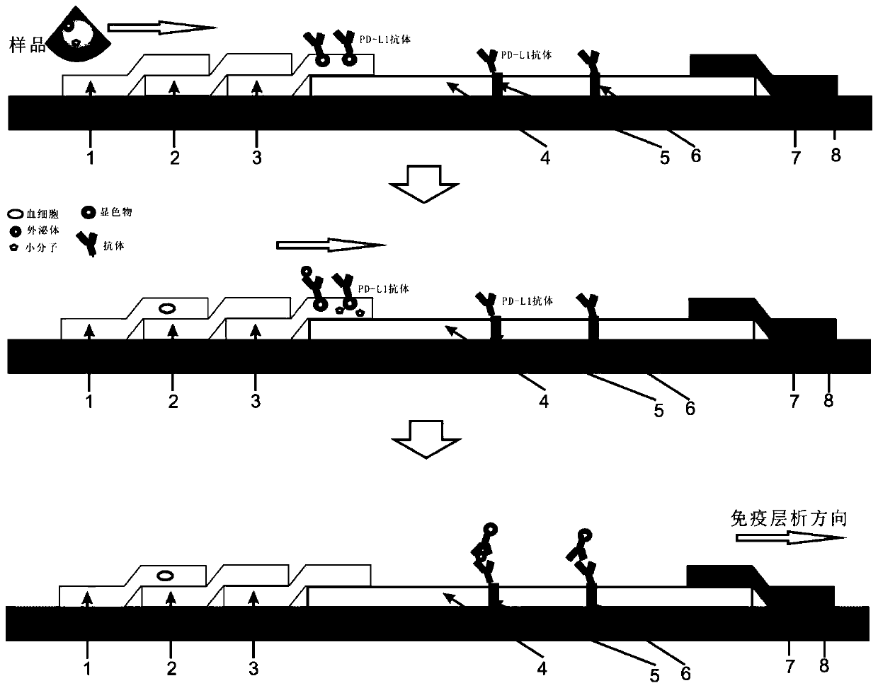
[0030] In vitro rapid detection platform and method for PD-L1 exosomes, including:
[0031] Sample primary filtration area 1: The sample primary filtration area is glass cellulose for primary filtration of whole blood, retaining most of the blood cells;
[0032] Nanofiltration area 2: The nanofiltration area 2 is connected to the sample primary filtration area 1; the nanofiltration area 2 is a nano-hydrophilic filter membrane, which realizes the further interception of residual blood cells in the previous step and at the same time realizes the separation of exosomes; nanofiltration The diameter of the membrane is 150 nm.
[0033] Chromogenic material pad area 3: the chromogenic material pad area 3 is connected to the nanofiltration area 2; the chromogenic material pad area 3 is used to fix the chromogenic material-labeled antibody, and can make the chromogenic material-labeled antibody and the sample complex dissolved; the chromogenic substance-labeled antibody is used to spe...
Embodiment 2
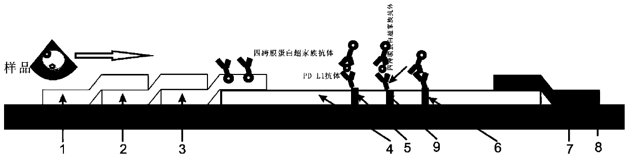
[0038] In vitro rapid detection platform and method for PD-L1 exosomes, including:
[0039] Sample primary filtration area 1: The sample primary filtration area is glass cellulose for primary filtration of whole blood, retaining most of the blood cells;
[0040]Nanofiltration area 2: The nanofiltration area 2 is connected to the sample primary filtration area 1; the nanofiltration area 2 is a nano-hydrophilic filter membrane, which realizes the further interception of residual blood cells in the previous step and at the same time realizes the separation of exosomes; nanofiltration The diameter of the membrane is 220 nm.
[0041] Chromogenic material pad area 3: the chromogenic material pad area 3 is connected to the nanofiltration area 2; the chromogenic material pad area 3 is used to fix the chromogenic material-labeled antibody, and can make the chromogenic material-labeled antibody and the sample complex dissolved; the chromogenic substance-labeled antibody is used to spec...
Embodiment 3
[0045] In vitro rapid detection platform and method for PD-L1 exosomes, including:
[0046] Sample primary filtration area 1: The sample primary filtration area is glass cellulose for primary filtration of whole blood, retaining most of the blood cells;
[0047] Nanofiltration area 2: The nanofiltration area 2 is connected to the sample primary filtration area 1; the nanofiltration area 2 is a nano-hydrophilic filter membrane, which realizes the further interception of residual blood cells in the previous step and at the same time realizes the separation of exosomes; nanofiltration The diameter of the membrane is 250 nm.
[0048] Chromogenic material pad area 3: the chromogenic material pad area 3 is connected to the nanofiltration area 2; the chromogenic material pad area 3 is used to fix the chromogenic material-labeled antibody, and can make the chromogenic material-labeled antibody and the sample complex dissolved; the chromogenic substance-labeled antibody is used to spe...
PUM
| Property | Measurement | Unit |
|---|---|---|
| diameter | aaaaa | aaaaa |
| diameter | aaaaa | aaaaa |
Abstract
Description
Claims
Application Information
 - R&D
- Intellectual Property
- Life Sciences
- Materials
- Tech Scout
- Unparalleled Data Quality
- Higher Quality Content
- 60% Fewer Hallucinations
Browse by: Latest US Patents, China's latest patents, Technical Efficacy Thesaurus, Application Domain, Technology Topic, Popular Technical Reports.
© 2025 PatSnap. All rights reserved.Legal|Privacy policy|Modern Slavery Act Transparency Statement|Sitemap|About US| Contact US: help@patsnap.com