Glycoprotein multi-charge isomer N-glycan structure evaluation method
A technology of charge isomerization and glycoprotein, applied in the field of protein medicine and bioengineering, can solve the problems of decreased cytotoxic activity, affecting cytotoxicity, reducing affinity, etc., and achieve the effect of improving clinical application, good reference significance, and convenient operation
- Summary
- Abstract
- Description
- Claims
- Application Information
AI Technical Summary
Problems solved by technology
Method used
Image
Examples
Embodiment 1
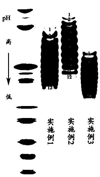
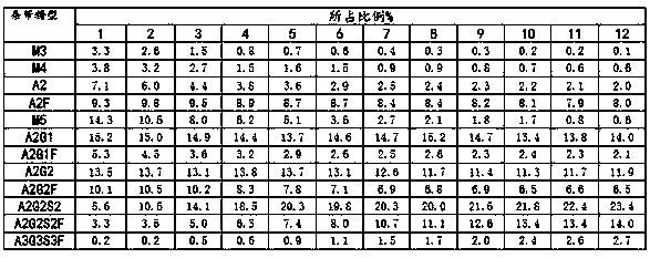
[0028] Example 1. Structural analysis of N-sugar chains of charge variants of recombinant human VEGFR-Fc fusion protein
[0029] (1) IEF separation charge heterogeneity
[0030] Take 1mg of recombinant human VEGFR-Fc fusion protein, use 10kD, 0.5ml ultrafiltration tube to replace liquid by ultrafiltration into 100μl ultrapure water, and quantify the protein concentration with a microplate reader. Take an appropriate amount of ultrafiltered sample and mix it with 2×sample buffer in an equal volume before loading the sample. The protein loading amount is 100 μg / band, and the gel used is pH3-10 IEF gel. Electrophoresis conditions were as follows.
[0031] Table 1 IEF electrophoresis voltage settings
[0032]
[0033] After electrophoresis, the gel was rinsed twice with ultrapure water, then soaked in 12% trichloroacetic acid fixative solution for fixation, and shaken at room temperature for 30 min. After that, the gel was washed 3 times with ultrapure water, and an appropri...
Embodiment 2
[0069] Example 2, High pI recombinant human VEGFR-Fc fusion protein charge variant N-sugar chain structure analysis
[0070] (1) IEF separation charge heterogeneity
[0071] Take 1mg of high pI recombinant human VEGFR-Fc fusion protein, use 10kD, 0.5ml ultrafiltration tube to replace liquid by ultrafiltration into 100μl ultrapure water, and quantify the protein concentration with a microplate reader. Take an appropriate amount of ultrafiltered sample and mix it with 2×sample buffer in an equal volume before loading the sample. The protein loading amount is 100 μg / band, and the gel used is pH3-10 IEF gel. Electrophoresis conditions were as follows.
[0072] Table 1 IEF electrophoresis voltage settings
[0073]
[0074] After electrophoresis, the gel was rinsed twice with ultrapure water, then soaked in 12% trichloroacetic acid fixative solution for fixation, and shaken at room temperature for 30 min. After washing the gel three times with ultrapure water, add an appropria...
Embodiment 3
[0103] Example 3, Low pI Recombinant Human VEGFR-Fc Fusion Protein Charge Isomer N Sugar Chain Structure Analysis
[0104] (1) IEF separation charge heterogeneity
[0105] Take 1 mg of the low pI recombinant human VEGFR-Fc fusion protein, use a 10kD, 0.5ml ultrafiltration tube to replace the medium by ultrafiltration into 100 μl ultrapure water, and quantify the protein concentration with a microplate reader. Take an appropriate amount of ultrafiltered sample and mix it with 2×sample buffer in an equal volume before loading the sample. The protein loading amount is 100 μg / band, and the gel used is pH3-10 IEF gel. Electrophoresis conditions were as follows.
[0106] Table 1 IEF electrophoresis voltage settings
[0107]
[0108] After electrophoresis, the gel was rinsed twice with ultrapure water, then soaked in 12% trichloroacetic acid fixative solution for fixation, and shaken at room temperature for 30 min. After washing the gel three times with ultrapure water, add an ...
PUM
 Abstract
Description
Claims
Application Information
 - R&D
- Intellectual Property
- Life Sciences
- Materials
- Tech Scout
- Unparalleled Data Quality
- Higher Quality Content
- 60% Fewer Hallucinations
Browse by: Latest US Patents, China's latest patents, Technical Efficacy Thesaurus, Application Domain, Technology Topic, Popular Technical Reports.
© 2025 PatSnap. All rights reserved.Legal|Privacy policy|Modern Slavery Act Transparency Statement|Sitemap|About US| Contact US: help@patsnap.com


