Method for in vitro proliferation of chondrocytes

A technology for chondrocytes and in vitro expansion, applied in the direction of bone/connective tissue cells, animal cells, vertebrate cells, etc., can solve the problems of ineffective application, selection of collection sites and limited collection volume, etc.

Pending Publication Date: 2019-07-16
THE SECOND PEOPLES HOSPITAL OF SHENZHEN
View PDF6 Cites 1 Cited by
  • Summary
  • Abstract
  • Description
  • Claims
  • Application Information

AI Technical Summary

Problems solved by technology

Including the method of transplanting cartilage tissue collected from other parts of the autologous body to the defect site for trea

Method used


Image

  • Method for in vitro proliferation of chondrocytes
  • Method for in vitro proliferation of chondrocytes
  • Method for in vitro proliferation of chondrocytes

Examples


Embodiment approach

[0087] According to a preferred embodiment of the present invention, the extract of Rhizoma Rhizoma Rhizoma and Rhizoma Drynariae extract are obtained as follows:

[0088] Step a, take 1 to 5 g, for example 2 g, of Rhizoma Rhizoma and Rhizoma Rhizoma Drynaria respectively, and then wash them with triple distilled water;

[0089] Step b. Soak in 200-800mL triple-distilled water, such as 400mL triple-distilled water, for 0.2-2h, such as 1h, and then simmer for 0.1-1h, such as 0.5h;

[0090] Step c, filtering respectively to obtain the Rhizoma Rhizoma Extract and Rhizoma Rhizoma Drynaria Extract.

[0091] According to a preferred embodiment of the present invention, in step 3, three-dimensional cell culture is used for subculture of cells.

[0092] In a further preferred embodiment, in step 3, the cells are subcultured using the polymer as a three-dimensional scaffold.

[0093] In a further preferred embodiment, in step 3, the cells are subcultured with polyε-caprolactone-b-pol...
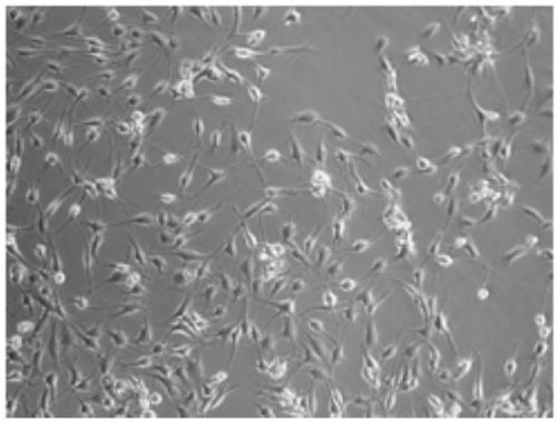
Embodiment 1

[0113] Chopped cartilage into 1.0mm 3 Size, washed 3 times with Ham's F12 medium (containing 10mmol / L HEPES buffer, 70μmol / L gentamicin sulfate, 22μmol / L amphotericin B, 300μmol / L ascorbic acid);

[0114] Put it into 9 mL of digestion medium (containing 1.5 mg / mL type II collagenase prepared in PBS) for digestion at 37 ° C for 15 hours, and then filter through a nylon mesh with a diameter of 40 μm;

[0115] Centrifuge at a speed of 1400 rpm, collect the cell pellet, and obtain chondrocytes;

[0116] 2500 pieces / cm 2 The cell density was seeded at 50cm 2 In the cell culture bottle, let stand for 2 days, wherein, the cell culture bottle is placed in the cell culture box (37 ℃, 5.0% CO 2 ), according to the GMP international quality control technical standard, the chondrocytes are placed in air cleanliness of 10000 grades, under the condition of 100 grades of biological safety cabinets for expansion and culture;

[0117] Carry out in DMEM medium (containing 8% human serum, 30...
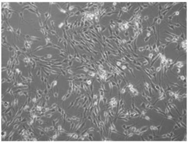
Embodiment 2

[0121] Chopped cartilage into 1.2mm 3 Size, washed 3 times with Ham's F12 medium (containing 15mmol / L HEPES buffer, 60μmol / L gentamicin sulfate, 25μmol / L amphotericin B, 250μmol / L ascorbic acid);

[0122] Put it into 10mL digestion medium (containing 2mg / mL type II collagenase prepared in PBS) and digest at 37°C for 13 hours, then filter through a nylon mesh with a diameter of 40μm;

[0123] Carry out centrifugation at a speed of 1300 rpm, collect cell pellets, and obtain chondrocytes;

[0124] 3500 pieces / cm 2 The cell density was seeded at 50cm 2 In the cell culture bottle, let stand for 2.5 days, wherein, the cell culture bottle is placed in the cell culture box (37 ℃, 5.0% CO 2 ), according to the GMP international quality control technical standard, the chondrocytes are placed in air cleanliness of 10000 grades, under the condition of 100 grades of biological safety cabinets for expansion and culture;

[0125] Cells were cultured in DMEM medium (containing 10% human s...


PUM


Abstract

The present invention discloses a method for in vitro proliferation of chondrocytes. The method comprises primary cell culture and cell passage culture. Growth factors of bFGF, PDGF, etc. are added ina primary culture medium to promote the primary cell culture, the growth factors of the bFGF, the PDGF, IGF-I, etc. having synergistic effects are added in a passage culture medium, so that the cellnumber is proliferated and at the same time cell hypertrophy can be effectively avoided; and at the same time, primary cells are subjected to a two-dimensional culture to enable the chondrocytes to beappropriately proliferated, and besides, preferably passage cells are subjected to a three-dimensional culture to restore phenotypes of degenerated cartilage tissues, so that the method proliferatesthe number of the chondrocytes and can also main the normal tissue phenotypes.

Description

technical field [0001] The invention belongs to the field of cell culture, in particular to in vitro culture and expansion of chondrocytes. Background technique [0002] Chondrocytes are the only cell type in articular cartilage, and they are terminal cells that lack the ability to proliferate in the internal environment of synovial fluid. In addition, articular cartilage has no blood supply, and once damaged, it lacks the ability to regenerate and repair. It includes the method of transplanting cartilage tissue collected from other parts of the autologous body to the defect site for treatment. This method has the problems of limited selection of collection sites and collection volume, and cannot be effectively applied. [0003] Therefore, there is an urgent need for an efficient method for in vitro expansion of chondrocytes. Contents of the invention [0004] In order to overcome the above-mentioned problems, the present inventors have carried out intensive research, add...

Claims


Application Information

Patent Timeline
IPC IPC(8): C12N5/077
CPCC12N5/0655C12N2501/105C12N2501/115C12N2501/135C12N2513/00
Inventor 段莉王大平李兴福徐晓陆伟熊建义
Owner THE SECOND PEOPLES HOSPITAL OF SHENZHEN
Why Eureka
Try Eureka