Thyroid nodule ultrasonic image classification method based on capsule network
A technology for thyroid nodules and ultrasound images, which is applied in image analysis, image enhancement, graphics and image conversion, etc. It can solve the problems of no translation of the model and loss of important information, etc., and achieve high accuracy and stable fluctuation of the accuracy change curve Effect
- Summary
- Abstract
- Description
- Claims
- Application Information
AI Technical Summary
Problems solved by technology
Method used
Image

Examples
Embodiment 1
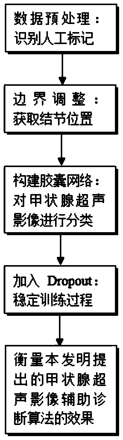
[0048] The embodiment of the present invention provides a method for classifying ultrasound images of thyroid nodules based on a capsule network, see figure 1 , the method includes the following steps:
[0049] 101: Preprocessing the ultrasound images of the thyroid gland to identify artificial markers in the ultrasound images;
[0050] 102: Obtain a rectangular frame for defining the position of the thyroid nodule through boundary adjustment;
[0051] 103: Construct a capsule network and apply it to the classification of ultrasound images of thyroid nodules, and adjust the structure of the capsule network;
[0052] 104: Add the Dropout method to the adjusted capsule network to stabilize the training process and improve the classification effect.
[0053] In one embodiment, the ultrasound image of the thyroid gland is preprocessed through step 101, and the specific steps are as follows:
[0054] In ultrasound images, a pair of "+" symbols (plus sign) are usually used to rep...
Embodiment 2
[0066] The scheme in embodiment 1 is further introduced below in conjunction with specific calculation formulas and examples, see the following description for details:
[0067] 201: Using image differentiation to identify artificial marks;
[0068] Among them, the Laplacian operator is a second-order differential operator. Applying a Laplacian operator L to the image R can obtain the second-order differential of the image R. In order to facilitate further processing, the second-order differential The obtained image is binarized and recorded as image G. The process is shown in formula (1), where η is the threshold used for image binarization, and the transformed image G is a binary image that only contains two gray values of 0 and 255. Among them, C=R×L.
[0069]
[0070] 202: The structure of the neural network used is shown in Table 1. After each convolutional layer, a ReLU is added as an activation function;
[0071] Table 1 Neural network structure for identifying ...
Embodiment 3
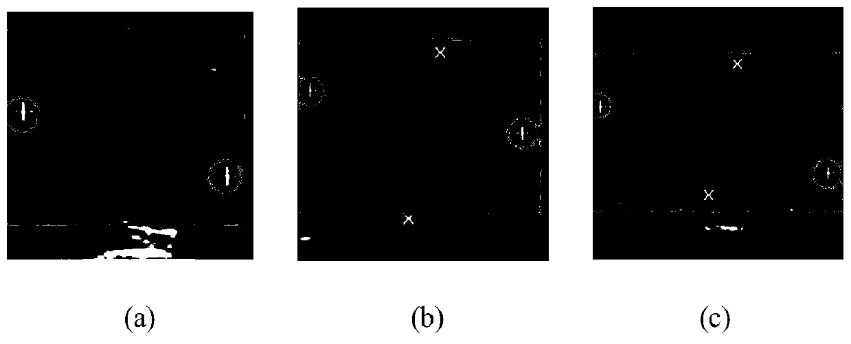
[0099] Combine below Figure 3-Figure 5 , and specific calculation formulas carry out feasibility verification to the scheme in embodiment 1 and 2, see the following description for details:
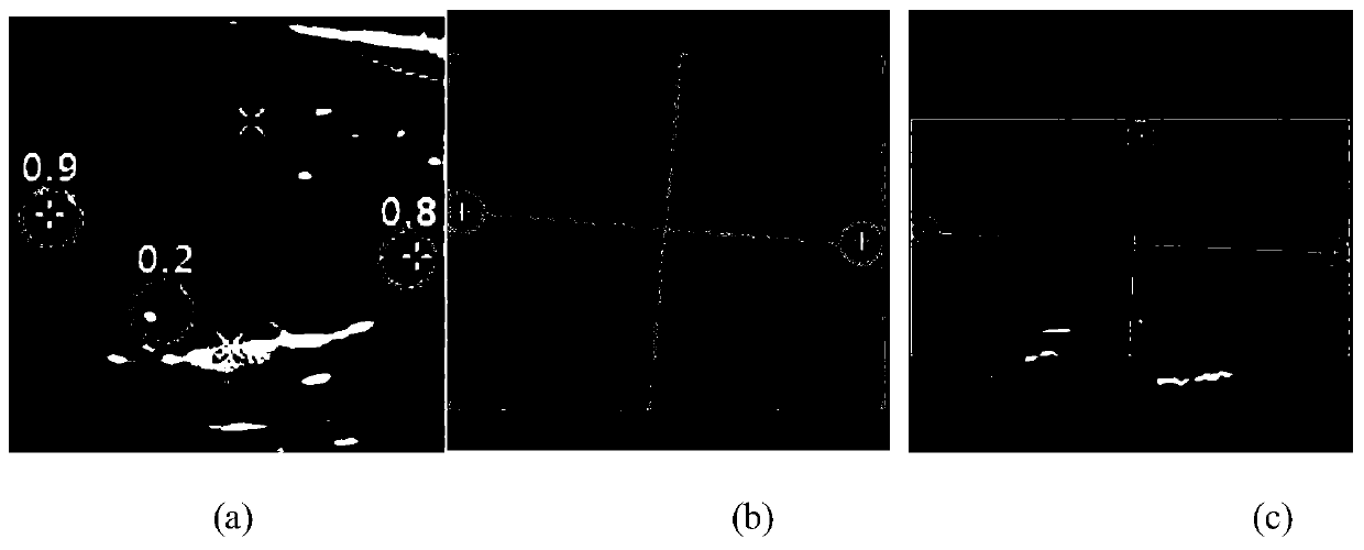
[0100] First of all, compared with other traditional methods, the method proposed in the embodiment of the present invention can accurately and quickly determine whether there is a manually marked symbol at a certain position in the ultrasound image, and give the category of the symbol and the probability of belonging to this category. The performance of our method compared with other methods is shown in Table 2. Combining the method of image preprocessing and border adjustment, it is possible to use a rectangular frame in the ultrasound image to locate the position of the thyroid nodule, such as image 3 shown. In the embodiment of the present invention, accuracy rate, precision rate, recall rate and F1 score are used as evaluation indicators, and the calculation formulas thereof are ...
PUM
 Abstract
Description
Claims
Application Information
 - R&D
- Intellectual Property
- Life Sciences
- Materials
- Tech Scout
- Unparalleled Data Quality
- Higher Quality Content
- 60% Fewer Hallucinations
Browse by: Latest US Patents, China's latest patents, Technical Efficacy Thesaurus, Application Domain, Technology Topic, Popular Technical Reports.
© 2025 PatSnap. All rights reserved.Legal|Privacy policy|Modern Slavery Act Transparency Statement|Sitemap|About US| Contact US: help@patsnap.com