Tooth jaw three-dimensional digital modeling method based on temporal-mandibular joint stability

A temporomandibular joint, three-dimensional digital technology, which is applied in the field of three-dimensional digital modeling of the teeth and jaws based on the stability of the temporomandibular joint, can solve the problem of high radiation risk, expensive, unaware of the importance of the close relationship between orthodontics, affecting orthodontics. effects, etc.

Active Publication Date: 2019-08-20
GUANGXI UNIVERSITY OF TECHNOLOGY
View PDF17 Cites 11 Cited by
  • Summary
  • Abstract
  • Description
  • Claims
  • Application Information

AI Technical Summary

Problems solved by technology

One reason is that the starting point of many orthodontics is to improve the facial appearance, without realizing the close connection between orthodontics and TMJ and the importance of TMJ stability in the long-term life of patients
The second reason is that there is no ready-made technical means to allow doctors to accurately consider the stability of TMJ in the process of orthodontic treatment planning
However, since the X-ray head image cannot give the three-dimensional shape and spatial relationship of the tissue structure, especially the stability of the TMJ structure cannot be judged from the two-dimensional image, so the existing models based on this technology have not included the state of the TMJ and its stability. Sexuality is included in the modeling considerations, which greatly affects the effect of orthodontics, causing long-term occlusal problems for patients
[0008] To sum up, the 3D digital model based on CT technology is difficult to be popularized in the foreseeable future due to its high radiation risk and high price, and cannot really help the diagnosis and treatment of clinical orthodontic surgery; The reconstructed 3D digital model has a basis for popularization on equipment, but none of them take the state of TMJ and its stability into consideration in modeling

Method used


Image

  • Tooth jaw three-dimensional digital modeling method based on temporal-mandibular joint stability

Examples


Embodiment Construction

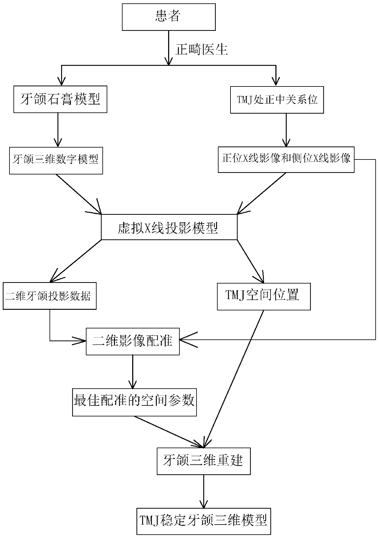
[0016] Such as figure 1 As shown, a kind of three-dimensional digital modeling method of dental jawbone based on temporomandibular joint stability of the present invention comprises the following steps:

[0017] Step 1: Take a plaster model of the patient's teeth and jaws, and use a 3D scanner to scan the plaster model to obtain a three-dimensional digital model of the teeth and jaws, or directly use an intraoral scanner to obtain a three-dimensional digital model of the teeth and jaws;

[0018] Step 2: The orthodontist adjusts and places the patient's temporomandibular joint (TMJ) in the median position, which means that the temporomandibular joint is in a stable state, and takes the patient's head frontal and lateral X-ray images;

[0019] Step 3: Refer to the spatial geometric relationship and imaging mechanism of the X-ray cephalometer, construct a virtual X-ray projection model, and its reference coordinate system, specifically refer to the X-ray cephalometer, construct a...


PUM


Abstract

The invention discloses a tooth jaw three-dimensional digital modeling method based on temporal-mandibular joint stability. The tooth jaw three-dimensional digital modeling method comprises the following steps of using a three-dimensional scanner for scanning to obtain a tooth jaw three-dimensional digital model of a patient; taking a positive position X-ray image and a lateral position X-ray image of the head of the patient at the median relation position; constructing a virtual X-ray projection model and a reference coordinate system; respectively adjusting positions and angles of upper andlower tooth jaws of the tooth jaw three-dimensional digital model in the virtual X-ray projection model, and calculating a two-dimensional X-ray projection image; registering the two-dimensional X-rayprojection images of the upper tooth jaw and the lower tooth jaw with an actual skull positive position X-ray image and an actual lateral position X-ray image, and recording parameters of the upper tooth jaw and the lower tooth jaw in a reference coordinate system during optimal registration; inversely solving spatial position information of the temporal-mandibular joint; reconstructing a dentaljaw three-dimensional digital model by using the upper and lower dental jaw three-dimensional digital model information during optimal registration; and further constructing a jaw occlusion motion model and a mechanical decomposition model of the three-dimensional digital model, and providing a virtual operation function and an operation prediction function according to the model.

Description

technical field [0001] The invention relates to the field of orthodontic digital technology, in particular to a three-dimensional digital modeling method for jawbone based on the stability of temporomandibular joints. Background technique [0002] Malocclusion is one of the high-incidence diseases in the department of stomatology, which will have adverse effects on personal oral health and appearance, and in severe cases, it will cause abnormal oral function. At present, orthodontics-orthognathic surgery is the main method for the treatment of malocclusion. [0003] An ideal orthodontic operation usually involves the following steps: 1. By observing the external manifestations of the patient’s malocclusion and asking about the relevant family history of the patient, a preliminary judgment can be made on the type of malocclusion and the genetic medical history; 2. The patient is supported by both hands Or put the patient's temporomandibular joint (TMJ) in the median position...

Claims


Application Information

Patent Timeline
Patent Type & Authority Applications(China)
IPC IPC(8): G06F17/50G06T7/33G06T17/00
CPCG06T17/00G06T7/344G06F30/23G06F2119/06Y02T90/00
Inventor 刘洪杨超蓝赠美刘晓晖王日凤李厚君
Owner GUANGXI UNIVERSITY OF TECHNOLOGY
Why Eureka
Try Eureka