Method for constructing biomimetic vascular net in large-volume tissue engineering tissue organ

A bionic blood vessel and tissue engineering technology, applied in the field of biomedical engineering, can solve problems such as simple network structure, supply barriers, lack of bile ducts, etc., and achieve the effect of mechanical strength

Active Publication Date: 2019-11-05
THE FIRST AFFILIATED HOSPITAL OF ARMY MEDICAL UNIV
View PDF8 Cites 7 Cited by
  • Summary
  • Abstract
  • Description
  • Claims
  • Application Information

AI Technical Summary

Problems solved by technology

For example, cells are seeded on a porous scaffold and soaked in the medium for culture, so that nutrients can reach the cells through the pores, but this method has certain limitations: as the cells grow, the pores in the scaffold will be filled by cells. The flow of nutrients is blocked, and the large-volume artificial tissue formed by this method is difficult to form the functional integration of cells in normal tissues due to the barrier of the scaffold material; in addition, some researchers use hydrogel materials that can induce blood vessel ingrowth However, this method still has the problem of deep support obstacles. When the blood vessels have not grown into the deep part, the deep cells will It has died due to lack of nutrients, and it is difficult to achieve long-term survival of real large-scale artificial tissues
The second product has the problems of short survival time, poor mechanical strength, simple vascular structure, single cells and simple network structure
These two products are limited by the incomplete vascular network system and in vitro support system, and both have the problem of limited size of the printed artificial liver tissue. After the size is limited, there is no way to simulate some functions of large-volume liver tissue, such as the lack of bile ducts. This structure lacks the function of bile transport
The products of some companies may form some pipelines by incorporating endothelial cells during printing, but these pipelines will not form a vascular network with complete functions of blood vessels such as penetration, transportation, and circulation. The functions are very different, it is not a bionic structure, and it is difficult to form a functional vascular network

Method used


Image

  • Method for constructing biomimetic vascular net in large-volume tissue engineering tissue organ
  • Method for constructing biomimetic vascular net in large-volume tissue engineering tissue organ

Examples


Embodiment Construction

[0037] In the present invention, the preparation of the blood vessel network in the artificial liver lobe containing bile duct is taken as an example to illustrate the method of the present invention.

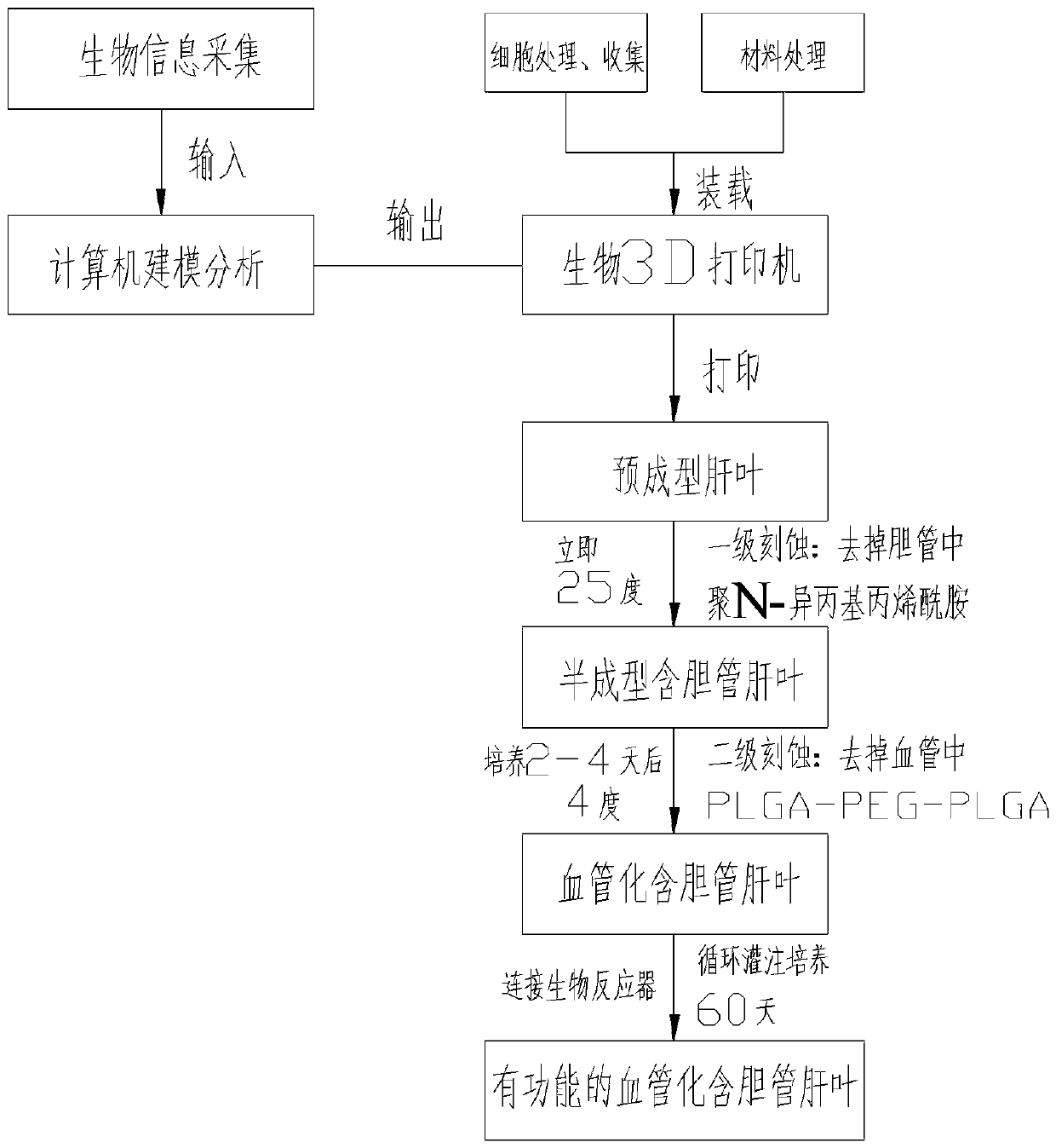
[0038] see figure 1 , this embodiment discloses a method for preparing an artificial liver lobe containing bile ducts based on bio-3D printing technology, comprising the following steps:

[0039] 1. Biological information collection and modeling:

[0040] 1) Personalized collection of three-dimensional data of the internal and external structures of the liver lobe containing bile ducts and the blood circulation network of normal people through CT, nuclear magnetic resonance and micro three-dimensional scanning technology;

[0041] 2) Input the collected biological information into the computer software, and imitate the actual tissue appearance and microenvironment to express it as a multi-material, multi-scale geometric model (the two ends of the vascular network are designed ...


PUM


Abstract

The invention discloses a method for constructing a biomimetic vascular net in a large-volume tissue engineering tissue organ. Firstly, biological information of a large-volume tissue of a human bodyis collected, and inputted into a computer for high biomimetic modeling; then, material separated printing and material-cell mixed printing output are performed with a hydrogel material and a varietyof amplified cells from the tissue by a high-precision biological 3D printer, wherein a hollow part in blood vessels is printed by a controllable degradable hydrogel; after all the hydrogel materialsare solidified, the controllable degradable hydrogel in the blood vessels are removed by means of specific enzymes or chelating reactions or temperature control or illumination and the like, and the vascular net in the large-volume artificial tissue is constructed. The circulation feeding of the vascular net and gas exchange in the large-volume artificial tissue are achieved by connection of mainblood vessels at two ends of the vascular net and a delivery pipe of a culture solution. The central feeding problem of the large-volume tissue is solved, so as to realize the long-term survival and biological function of the large-volume artificial tissue in vitro.

Description

technical field [0001] The invention belongs to biomedical engineering, and particularly relates to a bionic vascular network in a large-volume artificial tissue based on biological 3D printing and a preparation method thereof, that is, a vascular network with biological functions is prepared by using a biological 3D printing method to solve the problem of large-volume tissue Central feeding issues to achieve long-term survival and biological function of bulky artificial tissues in vitro. Background technique [0002] Existing artificial tissues are usually prepared by in vitro three-dimensional culture of cells, and only small-volume artificial tissues can be prepared. When the thickness of the artificial tissue exceeds 2 mm, it will cause deep tissue supply barriers, making it difficult for the artificial tissue to survive for a long time. In order to solve the problem of deep support barriers, researchers at home and abroad have carried out various explorations. For exa...

Claims


Application Information

Patent Timeline
Patent Type & Authority Applications(China)
IPC IPC(8): C12M3/00C12M1/36C12M1/34C12M1/04G06F17/50B29C64/112B29C64/379B29C64/386B33Y10/00B33Y40/00B33Y50/00
CPCC12M23/48C12M25/14C12M33/00C12M41/12C12M41/26C12M41/32C12M41/34C12M41/36C12M41/48B29C64/112B29C64/379B29C64/386B33Y10/00B33Y40/00B33Y50/00
Inventor 周强叶吉星甘翼搏李培涂兵罗磊赵晨欧阳斌张泽桐
Owner THE FIRST AFFILIATED HOSPITAL OF ARMY MEDICAL UNIV
Why Eureka
Try Eureka