
Structure, chip and use method for capturing circulating tumor cells of peripheral blood

A technology for peripheral blood circulation and tumor cells, which is applied in the structural field of capturing peripheral blood circulation tumor cells, can solve the problems of fast local flow velocity, uneven distribution of fluid pressure, and CTCs that cannot be captured, and achieve the effect of avoiding blockage

Pending Publication Date: 2019-12-13
SHENZHEN CHILDRENS HOSPITAL
View PDF6 Cites 2 Cited by
  • Summary
  • Abstract
  • Description
  • Claims
  • Application Information

AI Technical Summary

Problems solved by technology

[0004] Because the pore size of the filter membrane is uniform, and the diameter of many white blood cells is also greater than 8 microns, the filter membrane often has clogging problems when filtering peripheral blood. After local clogging, the fluid pressure will be unevenly distributed on the filter membrane. As a result, the local flow rate is too fast, and the CTCs in the peripheral blood cannot be intercepted, resulting in the failure of some CTCs to be captured

Method used

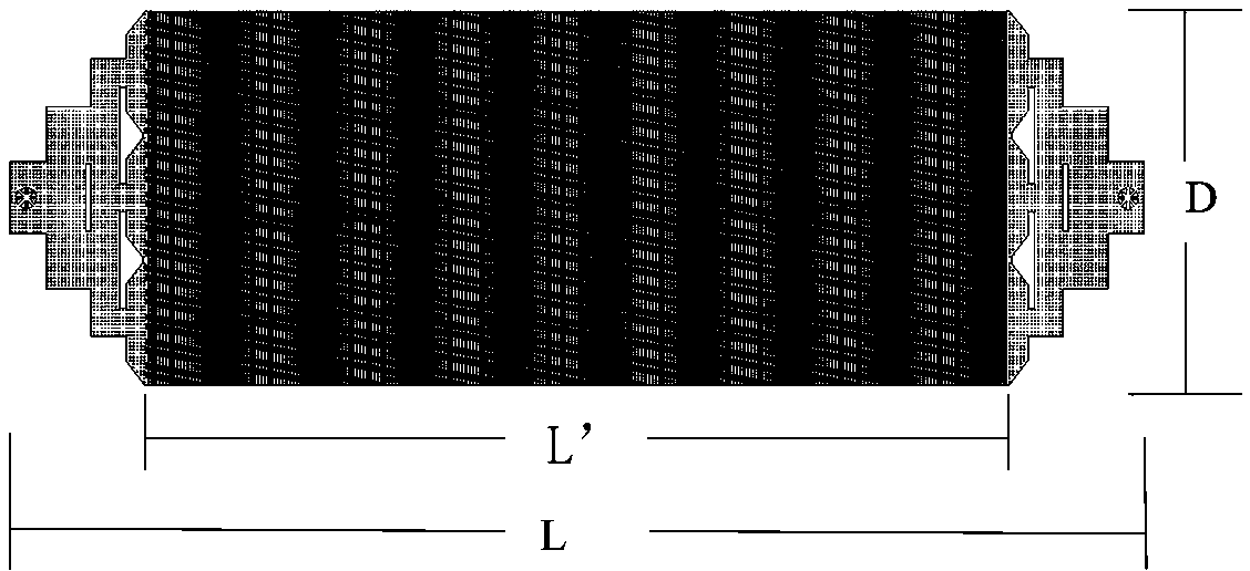
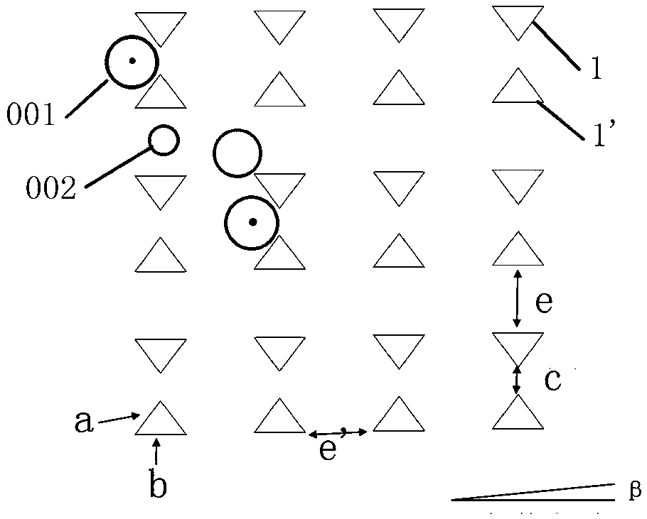

Image

  • Structure, chip and use method for capturing circulating tumor cells of peripheral blood
  • Structure, chip and use method for capturing circulating tumor cells of peripheral blood
  • Structure, chip and use method for capturing circulating tumor cells of peripheral blood

Examples


Embodiment 1

[0025] if figure 2 As shown, the length L of the chip is 47 mm, the width D is 15.5 mm, and the length L' of the intermediate cell capture region is 36 mm.

[0026] Such as image 3 As shown, the first capture block (1) and the second capture block (1') constituting the capture unit are both in the shape of a microcolumn (column) with a triangular cross-section. The two sides a of the triangle are 12.5 microns long and the bottom is b is 15 microns long, the interval c between the two opposite triangular micropillars in the capture unit is 8 microns, the longitudinal interval e and the transverse interval e' of the capture unit are both 20 microns, and the inclination angle β of the capture unit array is 1 degree, that is to say, The cells are arranged in a staggered arrangement so that if the first row does not capture the blood as it flows through, the second row in the staggered arrangement may capture it, and because there are many permutations, the tumor cells are captu...

Embodiment 2

[0031] if figure 2 As shown, the chip has a length of 47 mm, a width of 15.5 mm, and a length of 36 mm in the middle cell capture region. Such as Figure 5 As shown, the two sides of the triangular micropillars in the capture unit are 12.5 microns long, and the length of one side is 15 microns. The distance between the two opposite triangular microcolumns in the capture unit is 8 microns. All are 20 microns, the capture unit array has a tilt angle of 1 degree, the height of all microcolumns in the chip is 30 microns, and the total number of capture units in the chip is about 35*10 5 indivual.

[0032]A549 cells were stained with fluorescent dyes in advance, and about 1000 A549 cells labeled with fluorescent dyes were added to 2 ml of healthy human blood, and then 2 ml of blood was diluted to 10 ml with PBS (phosphate buffer saline) solution, and 10 One milliliter of blood is passed into the chip inlet through the syringe pump, and the flow rate of the solution is respectiv...


PUM

PropertyMeasurementUnit
Lengthaaaaaaaaaa
Widthaaaaaaaaaa
Heightaaaaaaaaaa

Abstract

The invention provides a structure, chip and use method for capturing circulating tumor cells of peripheral blood, and relates to the field of medical apparatuses. In order to increase the capturing rate of tumor cells and solve the problem that the chip is easy to block, the invention provides the following technical scheme of the structure for capturing circulating tumor cells of peripheral blood. The structure comprises a blood inlet, a waste blood outlet and a circulating tumor cell capturing region, wherein the circulating tumor cell capturing region comprises capturing units and releasechannels, and the capturing units are in misplaced arrangement in the blood flowing direction. The structure disclosed by the invention has the beneficial effects that when the peripheral blood pass through the chip / structure, large circulating tumor cells and a part of leucocyte (of which the diameter is 7-9 microns larger than a capturing interval) can be captured by the capturing units, the cells are stuck between the capturing units, other components in the blood can flow past the release channels on the two sides of the capturing units, blood blockage cannot be caused, and the purpose that the tumor cells in the blood can be captured in a higher efficiency manner is realized.

Description

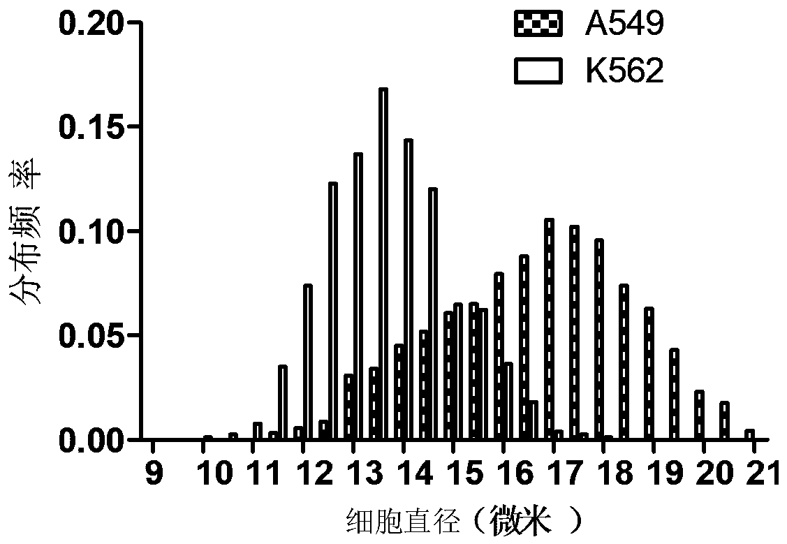
technical field [0001] The invention relates to the field of medical devices, in particular to a structure for capturing circulating tumor cells in peripheral blood, a chip and a method for using the same. Background technique [0002] Circulating tumor cells (CTCs) are tumor cells that come off from the primary tumor or metastases and enter the blood through the blood vessels or lymphatic system. CTCs derived from primary or metastatic tumors can not only indirectly reflect tumor characteristics, but also Real-time reflection of tumor development. CTCs are rare in peripheral blood, and there are only a few tumor cells among 100 million white blood cells and red blood cells. The separation and capture of CTCs is the basis for subsequent analysis (phenotype, genotype). [0003] The physical characteristics of tumor cells, such as size and density, are quite different from white blood cells and red blood cells. For example, the diameter of tumor cells is generally greater tha...

Claims


Application Information

Patent Timeline
IPC IPC(8): C12M1/00C12N5/09
CPCC12M47/04C12N5/0693Y02A50/30
Inventor 文飞球袁秀丽陈小文宋建明刘宗彬
Owner SHENZHEN CHILDRENS HOSPITAL
Learn More