Aortic valve semi-automatic segmentation method based on CTA dynamic image

A valve semi-automatic, dynamic image technology, applied in the field of medical image processing, can solve the problems of inability to automatically track, failure to present anatomical morphological feature information, and difficulty in showing the aortic valve structure, etc., achieving good temporal resolution and accuracy. The effect of spatial resolution

Active Publication Date: 2019-12-13
ARMY MEDICAL UNIV
View PDF6 Cites 9 Cited by
  • Summary
  • Abstract
  • Description
  • Claims
  • Application Information

AI Technical Summary

Problems solved by technology

However, the CTA imaging after the above CT scan is a regular transverse coronal and sagittal plane imaging, which is difficult to show the irregular membranous structure of the aortic valve, and the anatomical parameters it provides are limited to the static measurement of the aortic valve ring, and the dynamic measurement The difficulty lies in the fact that the heart is in constant motion, and the positions of its anatomical landmarks will also change in spa...

Method used


Image

  • Aortic valve semi-automatic segmentation method based on CTA dynamic image
  • Aortic valve semi-automatic segmentation method based on CTA dynamic image
  • Aortic valve semi-automatic segmentation method based on CTA dynamic image

Examples


Embodiment Construction

[0071] In order to make the purposes, technical solutions and advantages of the embodiments of the present application clearer, a clear and complete description will be made below in conjunction with the technical solutions in the embodiments of the present application. Obviously, the described embodiments are part of the embodiments of the present application, and Not all examples. Based on the embodiments of the present application, all other embodiments obtained by persons of ordinary skill in the art without making creative efforts belong to the protection scope of the present application.

[0072] The application is described in detail below in conjunction with the accompanying drawings:

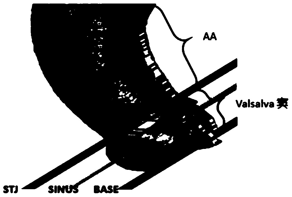
[0073] figure 1 The establishment image of the virtual annulus (Base) plane of the sinotubular junction (STJ) at the aortic root; figure 2 It is an image perpendicular to the plane of the aortic root sinus, including the aortic valve in the inner ring, the left coronary sinus on the ...


PUM


Abstract

The invention discloses an aortic valve semi-automatic segmentation method based on a CTA dynamic image, and the method comprises the steps: obtaining CT angiography image data under electrocardio gating, carrying out the division of 20 time phases, and carrying out the following steps for an image of each time phase point: step A, segmenting Valsalva sinus according to the upper and lower boundaries of the root of an aorta; and step B, segmenting the aortic valve from the Valsalva sinus. According to the method, the feature extraction range in the CTA image is expanded, so that more completeanatomical details are presented, and the working efficiency and safety during an operation are further improved.

Description

technical field [0001] The invention relates to the technical field of medical image processing, in particular to a semi-automatic segmentation method of an aortic valve based on a CTA dynamic image. Background technique [0002] The aortic valve is located at the root of the aorta and constitutes the physical boundary between the aorta and the left ventricle. It is in the morphological center of the heart and is also the hemodynamic center. Preventing aortic blood from returning to the left ventricle during diastole and allowing blood to flow from the left ventricle to the aorta during systole plays an important role in maintaining normal blood supply to the heart. [0003] The heart is a key organ of the human cardiovascular system. Its internal tissue structure is relatively complex, and the heart is constantly contracting. However, most of the image data of the heart are orthogonal two-dimensional plane images, and each plane is taken at a certain moment. For a certain ...

Claims


Application Information

Patent Timeline
IPC IPC(8): G06T7/00G06T7/11G06T7/136G06T7/155G06T7/187
CPCG06T7/0012G06T7/11G06T7/136G06T7/155G06T7/187G06T2207/10081G06T2207/30101
Inventor 李颖曲小龙陈伟陈永林李可谭立文张绍祥
Owner ARMY MEDICAL UNIV
Why Eureka
Try Eureka