Microwave thermoacoustic, optical acoustic and ultrasonic three-modal intestinal tissue imaging method and system

An ultrasonic imaging, three-modality technology, applied in the field of medical imaging, can solve the problems of intolerance of patients, difficult to enhance enhancement, long scanning time, etc., to achieve the effect of convenient operation

Inactive Publication Date: 2020-02-04
WEST CHINA HOSPITAL SICHUAN UNIV
View PDF12 Cites 5 Cited by
  • Summary
  • Abstract
  • Description
  • Claims
  • Application Information

AI Technical Summary

Problems solved by technology

[0002] Colonoscopy can better observe the lesions in the inner layer of the intestinal wall, as well as ulcers that reach deep into the muscular layer, mucosal exfoliation, or ulcers, etc., but it has limitations in the examination of intestinal wall structure changes and extraintestinal lesions. Microscopic examination is an invasive examination, which requires bowel preparation in advance, and the patient has a certain degree of pain during the examination process, which is difficult for some patients to tolerate, and is not suitable for cases of intestinal stenosis
[0003] X-ray barium meal examination of the intestinal tract can only show the lesions in the intestinal cavity, and due to the overlap of the intestinal anatomical structure, the thickness of the intestinal wall and the site of extraintestinal complications such as abdominal abscess cannot be displayed, which has great limitations
[0004] CT examination method is difficult to show mild inflammation of intestinal wall mucosa and superficial ulcer, and CT examination is radioactive, especially for young patients, so it is not suitable for long-term follow-up
[0005] Intestinal mucosal lesions displayed by MRI are not as good as X-ray barium meal examination, and artifacts caused by breathing or intestinal peristalsis are prone to occur due to the long scanning time
[0006] Ultrasonography is a non-invasive, non-radioactive, and convenient imaging method, but its operation is easily affected by the operator's technique and experience
Contrast-enhanced ultrasonography can better display the intestinal lumen and intestinal wall structure, but when the intestinal wall is too thin, it is difficult to enhance and display its enhancement

Method used


Image

  • Microwave thermoacoustic, optical acoustic and ultrasonic three-modal intestinal tissue imaging method and system
  • Microwave thermoacoustic, optical acoustic and ultrasonic three-modal intestinal tissue imaging method and system
  • Microwave thermoacoustic, optical acoustic and ultrasonic three-modal intestinal tissue imaging method and system

Examples


Embodiment 1

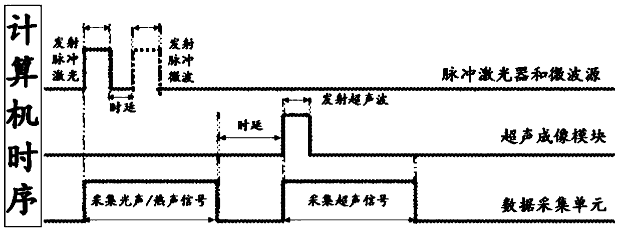
[0040] This embodiment provides a microwave thermoacoustic, photoacoustic and ultrasonic three-modal imaging method applied to intestinal inspection, including:

[0041] S1. Ultrasound imaging is performed on the area of ​​intestinal tissue to be imaged to obtain an ultrasonic image of intestinal tissue;

[0042] S2. Emit pulsed laser light and pulsed microwaves to the area of ​​intestinal tissue to be imaged, so that the area of ​​intestinal tissue to be imaged generates photoacoustic signals of intestinal tissue and thermoacoustic signals of intestinal tissue;

[0043] S3. Converting the photoacoustic signal of intestinal tissue and the thermoacoustic signal of intestinal tissue into a first electrical signal and a second electrical signal, respectively;

[0044] S4. Perform amplification and filtering processing on the first electrical signal and the second electrical signal;

[0045] S5. Converting the amplified and filtered first electrical signal and the second electric...

Embodiment 2

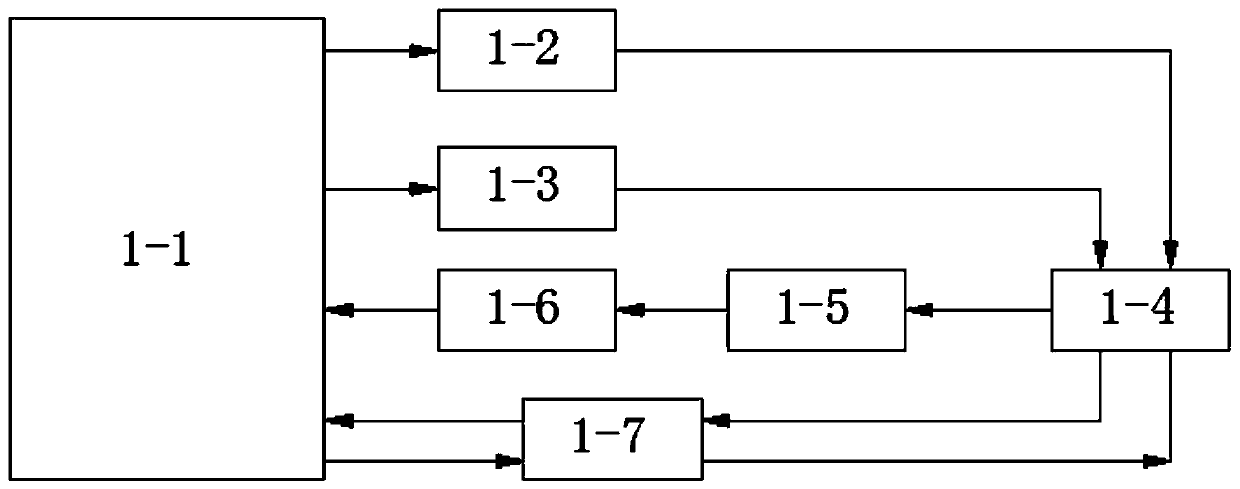
[0050] Please refer to figure 1 , this embodiment provides a microwave thermoacoustic, photoacoustic and ultrasonic three-modal imaging system applied to intestinal inspection, including:

[0051] Ultrasound imaging modules 1-7, which are used to acquire ultrasound images of intestinal tissue in areas to be imaged;

[0052]A pulsed laser 1-2, which is used to excite the area of ​​the intestinal tissue to be imaged to generate a photoacoustic signal of the intestinal tissue, which is an ultrasonic signal;

[0053] A pulsed microwave source 1-3, which is used to excite the area of ​​the intestinal tissue to be imaged to generate a thermoacoustic signal of the intestinal tissue, which is an ultrasonic signal;

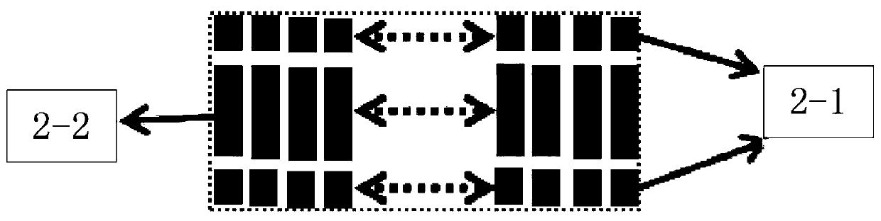
[0054] Ultrasonic transducers 1-4, which are used to receive photoacoustic signals of intestinal tissue and thermoacoustic signals of intestinal tissue, and convert both the photoacoustic signals of intestinal tissue and the thermoacoustic signals of intestinal tissue int...


PUM


Abstract

The invention discloses a microwave thermoacoustic, optical acoustic and ultrasonic three-modal intestinal tissue imaging method and system, and relates to a medical imaging technology. The microwavethermoacoustic, optical acoustic and ultrasonic three-modal intestinal tissue imaging method comprises the steps that intestinal tissue ultrasonic images are obtained, a pulse laser and a pulse microwave are emitted to a to-be-imaged area of intestinal tissue, thus intestinal tissue optical acoustic signals and intestinal tissue thermoacoustic signals are generated in the to-be-imaged area of theintestinal tissue and converted into electrical signals, through amplification, filtering, A/D conversion and image reconstruction, intestinal tissue optical acoustic images and intestinal tissue thermoacoustic images are obtained, and the intestinal tissue optical acoustic images subjected to color coding and the intestinal tissue thermoacoustic images subjected to color coding are superimposed on intestinal tissue ultrasonic images. The microwave thermoacoustic, optical acoustic and ultrasonic three-modal intestinal tissue imaging system comprises an ultrasonic imaging module, a pulsed laser, a pulse microwave source, a ultrasonic transducer, a signal processing unit, a data collecting unit and a computer. The imaging method is a non-invasive method, operation is convenient, patients donot have pain, and imaging of the intestinal tissue can be realized more comprehensively.

Description

technical field [0001] The invention relates to the technical field of medical imaging, in particular to a three-mode intestinal tissue imaging method and system of microwave thermoacoustic, photoacoustic and ultrasonic. Background technique [0002] Colonoscopy can better observe the lesions in the inner layer of the intestinal wall, as well as ulcers that reach deep into the muscular layer, mucosal exfoliation, or ulcers, etc., but it has limitations in the examination of intestinal wall structure changes and extraintestinal lesions. Endoscopic examination is an invasive examination that requires intestinal preparation in advance. During the examination process, the patient suffers a certain degree of pain, some patients cannot tolerate it, and it is not suitable for cases of intestinal stenosis. [0003] When X-ray barium meal is used to examine the intestinal tract, only the lesions in the intestinal cavity can be displayed, and due to the overlapping of intestinal anato...

Claims


Application Information

Patent Timeline
Patent Type & Authority Applications(China)
IPC IPC(8): A61B5/00A61B8/08A61B8/00
CPCA61B5/0093A61B5/0095A61B5/7225A61B8/085A61B8/5215
Inventor 李加伍
Owner WEST CHINA HOSPITAL SICHUAN UNIV
Why Eureka
Try Eureka