
Tumor surgical resection system

A technique for surgical resection and tumor resection, applied in the field of tumor surgical resection system, can solve problems such as difficult tumor boundary, and achieve the effects of avoiding errors, improving recognition accuracy, and improving accuracy

Active Publication Date: 2020-02-11
THE FIRST AFFILIATED HOSPITAL OF MEDICAL COLLEGE OF XIAN JIAOTONG UNIV
View PDF7 Cites 1 Cited by
  • Summary
  • Abstract
  • Description
  • Claims
  • Application Information

AI Technical Summary

Problems solved by technology

However, any resection method in the prior art has certain difficulties in determining the tumor boundary.

Method used


Image

  • Tumor surgical resection system
  • Tumor surgical resection system
  • Tumor surgical resection system

Examples


Embodiment 1

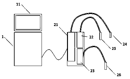
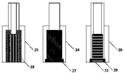
[0030] combine Figure 1-3 , a tumor surgical resection system, comprising a main control machine 1 and a drive cabinet 2; the main control machine 1 is provided with a display device 11, and an image collector 21, an infrared spectrometer 22, and a cutting laser source 23 are arranged in the drive cabinet 2; image acquisition Device 21 is connected with image acquisition probe 24, infrared spectrometer 22 is connected with infrared detection mark probe 25, and cutting laser source 23 is connected with laser cutting head 26; The image collector 21, the image collector 21 sends the image to the main controller 1 in real time, and displays it on the display device 11;

[0031] The infrared detection mark probe 25 can detect the Fourier transform infrared reflection spectrum of the tissue, and send the spectral data to the main controller 1, and the main controller 1 identifies the infrared spectrum to determine whether it is a tumor area; and the infrared detection mark probe 2...

Embodiment 2

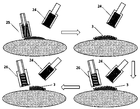
[0041] When the system works, under the illumination of the image acquisition probe 24, the infrared detection marker probe 25 is used to perform multiple infrared spectrum detections on the tissue determined as a tumor area and the tissue determined as a non-tumor area; and the detected infrared spectrum Send to the master controller 1;

[0042]The main control unit 1 constructs a tumor region recognition model by using the recognition model building block;

[0043] Use the infrared detection mark probe 25 to detect the infrared spectrum at the edge of the tumor area, and keep the infrared detection mark probe 25 and the tissue relatively fixed when collecting the infrared spectrum; the main controller 1 collects the unknown infrared spectrum collected by the infrared detection mark probe 25 Carry out analysis, and use the tumor area recognition model to determine whether it belongs to the tumor area;

[0044] If the collection area belongs to the tumor area, the infrared de...


PUM


Abstract

The present invention relates to a tumor surgical resection system. By utilizing an infrared spectrum detection means, infrared spectrum analysis can be performed firstly on a healthy area and a tumorarea, and an identification model is established in a host; then whether a boundary of the tumor area is the tumor area is identified according to the identification model and fluorescence labeling is also conducted; after the labeling, a laser cutting head is used for exciting fluorescence and then laser excision is conducted after the tumor area is confirmed; firstly the system can greatly improve identification accuracy of the tumor area and avoids errors of human eye identification; secondly, a focused focal point of the laser cutting head under a focusing working state is extremely small, so that cutting accuracy can be greatly improved; and the tumor area is labeled by using a fluorescent labeling mode, fluorescent identification is further conducted during cutting, the cutting accuracy is improved, labels fall off after cutting, and no influence is produced on healthy tissues.

Description

technical field [0001] The invention relates to the field of medical devices, in particular to a tumor surgical resection system. Background technique [0002] Tumor (tumour) refers to the neogrowth formed by the proliferation of local histiocytes under the action of various tumorigenic factors, because most of these neogrowths are space-occupying block-like protrusions, also known as neoplasms (neoplasm) . Tumor is one of the most important problems endangering human health. At present, about 1 / 3 of the people in the world suffer from cancer, and 1 / 4 of them die from cancer. [0003] Traditional tumor treatment methods are generally divided into radical tumor treatment and conservative treatment; the so-called radical tumor treatment generally involves surgical resection. However, during surgical resection, it is generally necessary to expand the scope of resection and remove suspicious tissues around the tumor. [0004] Application number: 201110037184.5 discloses a su...

Claims


Application Information

Patent Timeline
Patent Type & Authority Applications(China)
IPC IPC(8): A61B18/20A61B5/00
CPCA61B5/0071A61B5/0082A61B18/20A61B2018/00601A61B2018/20361
Inventor 刁冬梅程遥
Owner THE FIRST AFFILIATED HOSPITAL OF MEDICAL COLLEGE OF XIAN JIAOTONG UNIV
Learn More