Application of apoptotic body in preparation of products for promoting vascular regeneration of mammalian with tissue infarction

A technology of apoptotic bodies and mammals, applied in the field of regenerative medicine, to achieve the effect of promoting angiogenesis, stable quality, and good results

Inactive Publication Date: 2020-04-03
山东佰鸿干细胞生物技术有限公司
View PDF3 Cites 4 Cited by
  • Summary
  • Abstract
  • Description
  • Claims
  • Application Information

AI Technical Summary

Problems solved by technology

[0004] Acute myocardial infarction, known as the "number one killer" in developed countries, the existing treatment methods cannot fundamentally solve the problem of damaged myocardial cells and blood vessel regeneration and repair
It is inevitable to find more effective treatment methods and open up new treatment pathways. The pro-vascular effect of apoptotic bodies has not yet been publicly reported in the current technology.

Method used


Image

  • Application of apoptotic body in preparation of products for promoting vascular regeneration of mammalian with tissue infarction
  • Application of apoptotic body in preparation of products for promoting vascular regeneration of mammalian with tissue infarction
  • Application of apoptotic body in preparation of products for promoting vascular regeneration of mammalian with tissue infarction

Examples


Embodiment 1

[0070] Example 1: Isolation and purification of human umbilical cord mesenchymal stem cells

[0071] Discard the excess umbilical cord preservation solution in the obtained fresh umbilical cord, pour 75% alcohol for disinfection for three minutes, take out the umbilical cord, put it in physiological saline containing double antibodies and wash it repeatedly to remove the residual alcohol on the surface. Cut off the ligated part and discard it. Divide the remaining umbilical cord evenly into small pieces for cleaning, wash the residual blood repeatedly, remove the umbilical vein and umbilical artery, peel off the Wharton glue, and cut it to 1mm 3 The size of the organization block. According to the amount of tissue pieces, add an appropriate amount of conventional medium, mix evenly, spread it on a 15cm petri dish, and place it at 37°C, 5% CO 2 And cultivate in a humidity-saturated incubator for 24 hours, and add 8ml of culture solution to each culture dish. Replenish the flu...

Embodiment 2

[0072] Example 2: Induction of apoptosis of human umbilical cord mesenchymal stem cells and preparation of apoptotic bodies

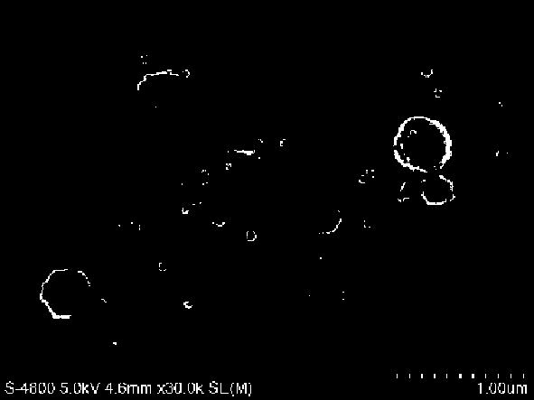
[0073] The human umbilical cord mesenchymal stem cells cultured to the 3rd-5th generation were taken at 2.6×10 6 The density of each well was inoculated into 6-well cell culture plates and placed at 37°C, 5% CO 2 The culture was continued under certain conditions, and when the cell confluency reached 80%-85%, a staurosporine solution was added for induction, and the concentration of the staurosporine ranged from 1 μm to 10 μm. After induction, the supernatant was collected. Supernatants from induced apoptotic cells were collected in sterile centrifuge tubes (approximately 15ml in approximately 50ml tubes).

[0074] Centrifuge the supernatant at 4°C, 400×g for 10 minutes, transfer the supernatant to remove cell debris; centrifuge the supernatant at 4°C, 2000×g for 10 minutes, transfer the supernatant to remove dead cells; The supernatant was further c...

Embodiment 3

[0076] Embodiment 3: MI model establishment, treatment and ultrasonic detection of SD rats

[0077] Select female SD rats weighing 180-200g, anesthetize with 1% pentobarbital sodium (40mg / kg) intraperitoneally, fix them on the experimental table in a supine position, connect them to an artificial respirator, and the respiratory rate is about 86 times / min. The tidal volume was 15ml, and the respiratory ratio was set at 1:1. Remove the hair on the left front chest, sterilize and spread the drape, incise the skin between the third and fourth ribs parasternal, bluntly separate the subcutaneous tissue and muscle, then cut the third rib, separate the heart with sutures, and place it at the lower edge of the left atrial appendage The left anterior descending coronary artery was ligated at 2 mm. After the ligation was completed correctly, the anterior wall of the left ventricle and the apex of the heart could be observed to be white. Then the chest cavity was sutured from the inside t...


PUM

PropertyMeasurementUnit
diameteraaaaaaaaaa
molecular weightaaaaaaaaaa

Abstract

The invention discloses an apoptotic body produced by inducing apoptosis of human umbilical cord mesenchymal stem cells in vitro and application thereof for promoting vascular regeneration of rats with myocardial infarction, and belongs to the field of regenerative medicine. At the same time, the invention also relates to a preparation method for obtaining the apoptotic body by inducing apoptosisof human umbilical cord mesenchymal stem cells in vitro. Experiments in vitro of the invention prove that the induction of apoptosis of umbilical cord mesenchymal stem cells can produce the apoptoticbody. At the same time, experiments in vivo prove that when the induced apoptotic body is used in the treatment of rats with myocardial infarction, the recovery of rat heart function and the vascularregeneration of damaged myocardium can be promoted. Therefore, the apoptotic body may become a potential effective agent for improving cardiac function after myocardial infarction.

Description

technical field [0001] The invention belongs to the field of regenerative medicine. In particular, the invention relates to the application of an apoptotic body in the preparation of a blood vessel regeneration product for promoting mammalian tissue infarction. Background technique [0002] Apoptosis is a highly regulated cell death process, unlike necrosis, which is traumatic death of cells. Apoptosis is a rational and aggressive physiological process that sacrifices specific cells for greater benefit to the normal physiological processes of the organism. Clearance of apoptotic cells has been a major research area of ​​apoptosis-related research. Efficient and timely clearance of apoptotic cells is critical to avoid immune responses to self-antigens and prevent perturbation of surrounding cells and tissues. Most cells undergoing apoptosis are eliminated by phagocytes in the form of small vesicles called apoptotic bodies (ABs). Compared with exosomes and microvesicles, A...

Claims


Application Information

Patent Timeline
Patent Type & Authority Applications(China)
IPC IPC(8): A61K35/51A61P9/10A61P11/00A61P1/00A61P21/00C12N5/0775
CPCA61K35/51A61P1/00A61P9/10A61P11/00A61P21/00C12N5/0668C12N2509/00
Inventor 蒲锋星
Owner 山东佰鸿干细胞生物技术有限公司
Why Eureka
Try Eureka