
Method for amplifying cone photoreceptors or rod photoreceptors by using dorsalization signal transmitter or ventralization signal transmitter

A technology of cone cells and rod cells, applied in the field of retinal tissue

Pending Publication Date: 2020-05-01
RIKEN +2
View PDF26 Cites 3 Cited by
  • Summary
  • Abstract
  • Description
  • Claims
  • Application Information

AI Technical Summary

Problems solved by technology

[0006] However, how dorsalized regions participate in the formation of rod-free regions in human or chicken retinal tissue is unknown

Method used


Image

  • Method for amplifying cone photoreceptors or rod photoreceptors by using dorsalization signal transmitter or ventralization signal transmitter
  • Method for amplifying cone photoreceptors or rod photoreceptors by using dorsalization signal transmitter or ventralization signal transmitter
  • Method for amplifying cone photoreceptors or rod photoreceptors by using dorsalization signal transmitter or ventralization signal transmitter

Examples


preparation example Construction

[0123]Preparation of target vectors for homologous recombination target genes and efficient sorting of homologous recombination can be carried out according to the following methods: Gene Targeting, A Practical Approach (gene targeting, practical method), IRLPress at Oxford University Press (1993); Biomanual Series 8, Gene Targeting, Making of Mutant Mouse using ES cell (gene targeting, using ES cells to produce mutant mice), YODOSHA CO., LTD. (1995); etc. Either a substitution type or an insertion type can be used for the targeting vector. As a sorting method, methods such as positive selection, promoter selection, negative selection, and polyadenylic acid (polyA) selection can be used.

[0124] As a method for selecting a desired homologous recombinant from the sorted cell lines, DNA hybridization method (Southern hybridization method) to genomic DNA, PCR method, etc. are mentioned.

[0125] The "suspension culture" or "suspension culture method" in the present invention re...

Embodiment 1

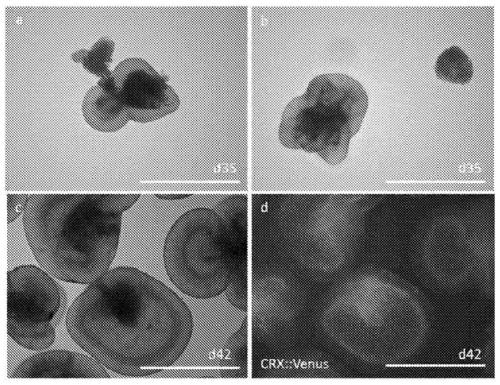
[0653] Example 1 (Preparation of Cell Aggregates Containing Retinal Tissue Using Human ES Cells and Excision Method of Retinal Tissue)

[0654] Human ES cells (derived from KhES-1; Nakano, T. et al. Cell Stem cell2012, 10(6), 771-785) knocked into CRX::Venus were based on Ueno, m. et al. PNAS 2006, 103 (25), 9554-9559" and "Watanabe, K. et al. Nat Biotech 2007, 25, 681-686". As for the culture medium for human ES cells, DMEM / F12 20% KSR (Knockout TM Serum replacement; Invitrogen), 0.1 mM 2-mercaptoethanol, 2 mM L-glutamine, 1x non-essential amino acids, 7.5 ng / mL bFGF medium.

[0655] Cell aggregates including retinal tissue were prepared by modifying a part of the method described in "Kuwahara et al. Nat Commun 2015, 19(6), 6286-". That is, after the previously cultured ES cells were dispersed into individual cells using TrypLE Express (Invitrogen), the individual dispersed human ES cells were separated into 9×10 cells per well. 3 Cells were suspended in 100 μL of serum-fr...

Embodiment 2

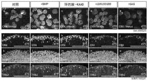
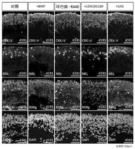
[0661] In the method described in Example 1, a dorsalization signal transduction substance (BMP4 or cyclopamine-KAAD) or a ventralization signal transduction substance (LDN193189 or SAG) was added as follows to prepare a retinoid-rich Aggregates of retinal tissue of cone or rod precursor cells.

[0662] [1] Control group ( figure 2 a, f, k, p); According to the method described in Example 1, culture was carried out without adding any substance until the 70th or 75th day after the start of the suspension culture.

[0663] [2]+BMP group ( figure 2 b, g, l, q); From the 22nd day to the end of the suspension culture, 0.15nM BMP4 was added, and cultured to the 70th day or the 75th day after the suspension culture was started.

[0664] [3]+Cyclopamine-KAAD group ( figure 2 c, h, m, r); from the 22nd day to the end of the suspension culture, 500 nM cyclopamine-KAAD was added, and cultured to the 70th or 75th day after the suspension culture started.

[0665] [4]+LDN193189 grou...


PUM

PropertyMeasurementUnit
Diameteraaaaaaaaaa

Abstract

The present invention addresses the problem of providing a cone photoreceptor precursor cell and / or a retinal tissue rich in cone photoreceptors, a rod photoreceptor precursor cell and / or a retinal tissue rich in rod photoreceptors, and a method for manufacturing the same. The present invention is i) a method for amplifying the ratio of cone photoreceptor precursor cells and cone photoreceptors inphotoreceptor precursor cells and photoreceptors included in a retinal tissue, the method including a step for culturing the retinal tissue from an initial development stage until a stage at which the occurrence of cone photoreceptor precursor cells is at maximum in a medium including a dorsalization signal transmitter at a concentration sufficient to suppress expression of a ventral marker; or ii) a method for amplifying the ratio of rod photoreceptor precursor cells and rod photoreceptors in photoreceptor precursor cells and photoreceptors included in a retinal tissue, the method includinga step for culturing the retinal tissue from an initial development stage until a stage at which the occurrence of rod photoreceptor precursor cells is at maximum in a medium including a dorsalizationsignal transmitter at a concentration sufficient to suppress expression of a ventral marker.

Description

technical field [0001] The present invention relates to retinal tissues rich in cone cells or rod cells and their preparation methods. Background technique [0002] Retinal tissues of mice, rats, humans, and chickens are all said to be determined by dorsalization signals and ventralization signals, but each has a unique structure depending on the species of animal. [0003] For example, in the retinas of humans and chickens, unlike mice and rats, there is an area with a high ratio of cone cells (cones) called the macula in the fundus (or central retina), and in the center There are only cone cells in the center (no rod cell area), especially in the macula in human retinal tissue, the closer to the center, the higher the content of LM cone cells, and the more peripheral, on the contrary the S cone The higher the percentage of cells. In addition, there is an area including S cone cells and rod cells outside the macula, and rod cells are densely packed in the outer edge of th...

Claims


Application Information

Patent Timeline
IPC IPC(8): C12N5/071A61K35/30A61L27/38A61P27/02
CPCA61K35/30A61P27/02C12N2501/155C12N2501/415C12N2501/41C12N5/0621C12N2506/02A61L2430/16A61L27/3604A61L27/3804A61L27/3834A61F2/14
Inventor 糠谷大树永乐元次木濑结子大西晓士高桥政代笹井芳树
Owner RIKEN
Learn More