Cell interpretation method and system based on FISH technology

A cell and technology technology, applied in the field of circulating tumor cell detection, can solve the problems of low throughput and low accuracy of FISH staining cells in the manual interpretation process, and achieve the effect of alleviating the low throughput and low accuracy of the manual interpretation process.

Pending Publication Date: 2020-05-19
ZHUHAI LIVZON CYNVENIO DIAGNOSTICS +1
View PDF5 Cites 12 Cited by
  • Summary
  • Abstract
  • Description
  • Claims
  • Application Information

AI Technical Summary

Problems solved by technology

[0005] In view of this, the object of the present invention is to provide a cell interpretation method and system based on FISH technology to alleviate the low accuracy and low throughput of manual interpretation process in the prior art in the process of automatic analysis and processing of FISH stained cells question

Method used


Image

  • Cell interpretation method and system based on FISH technology
  • Cell interpretation method and system based on FISH technology
  • Cell interpretation method and system based on FISH technology

Examples


Embodiment 1

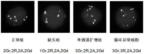
[0025] Auxiliary diagnostic test for benign and malignant pulmonary nodules includes automatic imaging and analysis of 10,000 FISH cells using a high magnification objective lens, and then interpretation of all automatically sorted cells. Based on the consideration of reducing the automatic imaging time and interpretation time, the present invention provides a cell interpretation method based on FISH technology.

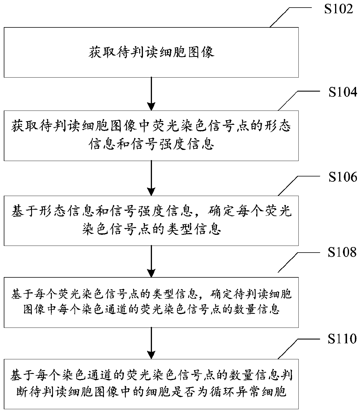
[0026] figure 1 It is a flow chart of a cell interpretation method based on FISH technology provided according to the embodiment of this aspect. Such as figure 1 As shown, the method specifically includes the following steps:

[0027] Step S102, acquiring the image of the cell to be interpreted; the image of the cell to be interpreted is the cell image after staining the chromosome of the cell to be interpreted by the in situ fluorescence hybridization technique, and the image of the cell to be interpreted contains fluorescent staining signal points of multiple sta...

Embodiment 2

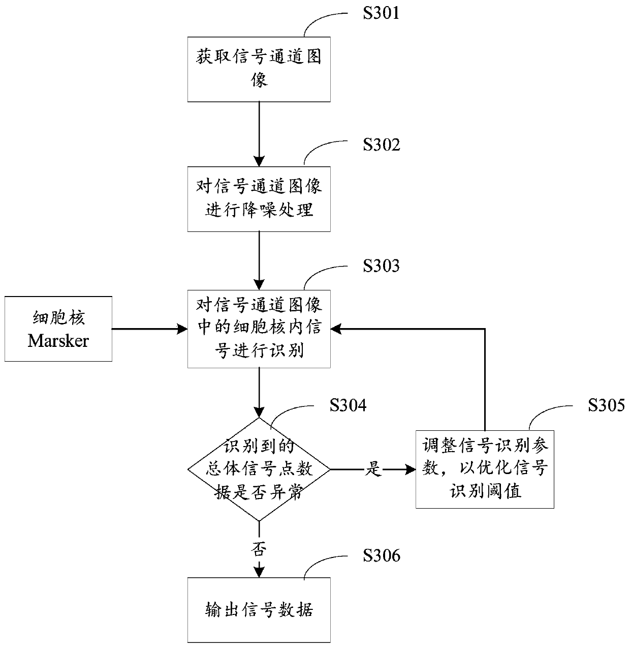
[0075] Figure 4 It is a schematic diagram of a cell interpretation system based on FISH technology provided according to an embodiment of the present invention, such as Figure 4 As shown, the system includes: a first acquisition module 10 , a second acquisition module 20 , a first determination module 30 , a second determination module 40 and an interpretation module 50 .

[0076] Specifically, the first acquisition module 10 is configured to acquire an image of a cell to be interpreted; the image of a cell to be interpreted is a cell image obtained by staining the chromosome of the cell to be interpreted by the in situ fluorescence hybridization technique, and the image of the cell to be interpreted includes Fluorescent staining signal points for multiple staining channels.

[0077] The second acquiring module 20 is configured to acquire the morphological information and signal intensity information of the fluorescent staining signal points in the cell image to be interpre...


PUM


Abstract

The invention provides a cell interpretation method and system based on an FISH technology. The cell interpretation method comprises steps of acquiring a to-be-interpreted cell image; acquiring the morphological information and the signal intensity information of fluorescence staining signal points in the to-be-interpreted cell image; determining the type information of each fluorescence stainingsignal point based on the morphological information and the signal intensity information; wherein the type information comprises any one of a normal signal point, a strong signal point, a weak signalpoint, a split signal point and a rod-shaped signal point; based on the type information of each fluorescent staining signal point, determining the quantity information of the fluorescent staining signal points of each staining channel in the to-be-interpreted cell image; and judging whether cells in the to-be-interpreted cell image are circulating abnormal cells or not based on the quantity information of the fluorescent staining signal points of each staining channel. The method is advantaged in that technical problems that in the prior art, in the automatic analysis and treatment process ofthe FISH stained cells, accuracy is low, and the flux in the manual interpretation process is low are solved.

Description

technical field [0001] The present invention relates to the technical field of detecting circulating tumor cells, in particular to a cell interpretation method and system based on FISH technology. Background technique [0002] Fluorescence in situ hybridization (FISH) is an important non-radioactive in situ hybridization technique, which appeared in the genetics experiment technique in the late 1970s. According to the principle of complementary base pairing, the probe with fluorescent substance is combined with the target DNA by special means, and finally the position of the target DNA can be directly observed with a fluorescence microscope. [0003] Direct observation of target DNA requires the use of high magnification microscopes. The high magnification limits the number of observed cells in the field of view. In addition, a high-magnification lens with a shallow depth of field is not enough to collect the fluorescent signals of all the target DNA in the nucleus in one ...

Claims


Application Information

Patent Timeline
Patent Type & Authority Applications(China)
IPC IPC(8): G01N21/64
CPCG01N21/6402G01N21/6486
Inventor 叶莘范献军陈燕慈黄萌周燕玲
Owner ZHUHAI LIVZON CYNVENIO DIAGNOSTICS
Why Eureka
Try Eureka