
PET reconstruction method based on anatomical prior information

A technology of prior information and anatomical features, applied in the field of medical PET imaging, can solve the problems of image blur, poor quantitative analysis effect, restricting the PET system, etc., to achieve good reconstruction quality, improve image signal-to-noise ratio and quantitative analysis effect.

Pending Publication Date: 2020-07-10
广州互云医院管理有限公司
View PDF0 Cites 1 Cited by
  • Summary
  • Abstract
  • Description
  • Claims
  • Application Information

AI Technical Summary

Problems solved by technology

The spatial resolution of PET is one of the core indicators of the PET system. Some physical characteristics of positrons and the limitations of the data acquisition mechanism lead to an inevitable reduction in spatial resolution, which is manifested as blurred images on the image, which restricts the PET system. In some applications that require high resolution, such as early tumor discovery, brain functional imaging, etc.
[0003] The traditional PET image reconstruction method has high image noise, resulting in poor quantitative analysis and affecting image diagnosis. In order to solve the above technical problems, a new technical solution is proposed.

Method used


Image

  • PET reconstruction method based on anatomical prior information
  • PET reconstruction method based on anatomical prior information

Examples


Embodiment Construction

[0018] The following will clearly and completely describe the technical solutions in the embodiments of the present invention with reference to the accompanying drawings in the embodiments of the present invention. Obviously, the described embodiments are only some, not all, embodiments of the present invention. Based on the embodiments of the present invention, all other embodiments obtained by persons of ordinary skill in the art without making creative efforts belong to the protection scope of the present invention.

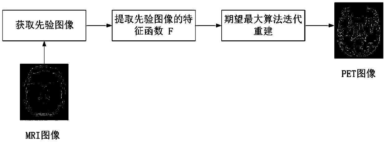
[0019] See Figures, a method for PET reconstruction based on anatomical prior information, which consists of the following steps:

[0020] Step 1. Obtain a priori image;

[0021] Step 2, extracting a function F representing anatomical features in a high-dimensional space;

[0022] Step 3. Select the radial Gaussian kernel function;

[0023] Step 4, adding the kernel method to the iterative reconstruction framework of the expected maximum algorithm, which is ...


PUM


Abstract

A PET reconstruction method based on anatomical prior information relates to the medical PET imaging technology, and comprises the following steps: 1, obtaining a prior image; 2, extracting a functionF representing anatomical features in a high-dimensional space; 3, selecting a radial Gaussian kernel function; 4, adding a kernel method into the expectation maximization algorithm iterative reconstruction framework to represent the PET image in the reconstruction process; 5, finally obtaining a reconstruction result, adding the extracted priori information of the anatomical image with high spatial resolution into a kernel method reconstruction process based on machine learning, obtaining better PET image reconstruction quality, and improving an image signal-to-noise ratio and a quantitativeanalysis effect.

Description

technical field [0001] The invention relates to medical PET imaging technology, in particular to a PET reconstruction method based on anatomical prior information. Background technique [0002] Positron Emission Computed Tomography (PET), as a non-interventional tool for quantitatively studying functional activity in vivo, is being more and more widely used in clinical diagnosis, especially early diagnosis of diseases. The spatial resolution of PET is one of the core indicators of the PET system. Some physical characteristics of positrons and the limitations of the data acquisition mechanism lead to an inevitable reduction in spatial resolution, which is manifested as blurred images on the image, which restricts the PET system. In some applications that require high resolution, such as early tumor discovery, brain functional imaging, etc. [0003] The traditional PET image reconstruction method has high image noise, resulting in poor quantitative analysis and affecting imag...

Claims


Application Information

Patent Timeline
Patent Type & Authority Applications(China)
IPC IPC(8): G06T11/00
CPCG06T11/003
Inventor 刘新黄志炜李晓伟陈铭湘
Owner 广州互云医院管理有限公司
Learn More