Retinal angiography optical imaging system and method

A technology of retinal blood vessels and contrast imaging, which is applied in the field of optics and can solve problems such as complex operations and allergies

Active Publication Date: 2020-08-14
宁波蓝明信息科技有限公司
View PDF7 Cites 0 Cited by
  • Summary
  • Abstract
  • Description
  • Claims
  • Application Information

AI Technical Summary

Problems solved by technology

However, the disadvantage of fluorescein angiography is that its imaging requires the injection of contrast agents, so this method is an invasive, complicated detection method and has risks such as allergies

Method used


Image

  • Retinal angiography optical imaging system and method
  • Retinal angiography optical imaging system and method
  • Retinal angiography optical imaging system and method

Examples


Embodiment Construction

[0010] Specific embodiments of the present invention will be described in detail below in conjunction with technical solutions and drawings.

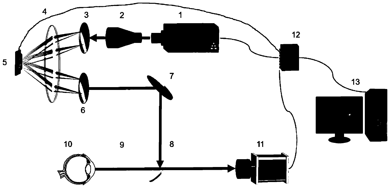
[0011] As shown in the figure, a retinal vascular optical contrast imaging system includes a white light source 1, a beam expander 2, a first grating 3, a first lens 4, a digital micromirror device 5, a second grating 6, a mirror 7, and a beam splitter Mirror 8, second lens 9, CCD camera 11, data acquisition card 12 and computer 13;

[0012] It consists of a white light source 1, a beam expander 2, a first grating 3, a first lens 4, a digital micromirror device 5, a second grating 6, a reflector 7, a beam splitter 8 and a second lens 9 for illuminating the eye to be inspected 10 lighting units;

[0013] An imaging unit for imaging the subject's eye 10 is composed of the second lens 9, the beam splitter 8 and the CCD camera 11;

[0014] A control unit for controlling the lighting unit and the imaging unit is composed of a digital micro...


PUM


Abstract

The invention discloses a retinal angiography optical imaging system and method, and belongs to the technical field of optics. The system comprises an illumination unit used for illuminating an examined eye, an imaging unit used for collecting retinal image information, and a control unit used for carrying out coding regulation and control on light emitted by a light source. According to the method, coding regulation and control are carried out on light entering the fundus on the aspect of wavelength through a coding matrix to form special illumination light sensitive to retinal blood vessels,the contrast of the retinal blood vessels is enhanced by collecting retinal images illuminated by the special illumination light, and the contrast ratio of the retinal blood vessels is improved. An optical means is used for replacing the specific selection process of a contrast agent for blood vessels in traditional fundus fluorescence angiography imaging, and a simple, noninvasive and high-contrast retinal angiography imaging method is achieved.

Description

technical field [0001] The invention belongs to the field of optics, and in particular relates to a retinal blood vessel optical contrast imaging system and method. Background technique [0002] Retinal blood vessels are blood vessels that can be observed non-invasively. Doctors can diagnose fundus-related diseases (such as age-related macular degeneration, diabetic retinopathy, glaucoma, etc.) The clinical treatment process can effectively prevent vision loss or blindness. [0003] In clinical practice, retinal blood vessels are usually imaged and observed by methods such as color fundus camera and fundus fluorescein angiography. Among them, the color fundus camera mainly images the surface of the fundus, and stores the condition of the fundus in the form of images. The color fundus camera imaging is fast and easy to operate, which greatly facilitates the routine examination of ophthalmology. However, the contrast of the fundus images collected by it is low, and it is of...

Claims


Application Information

Patent Timeline
Patent Type & Authority Applications(China)
IPC IPC(8): A61B3/14A61B3/12
CPCA61B3/14A61B3/0016A61B3/12A61B3/0008
Inventor 陈硕孔令敏佟萌萌
Owner 宁波蓝明信息科技有限公司
Why Eureka
Try Eureka