DICOM image processing method and system

An image processing and image technology, applied in the field of image processing, can solve the problem of wasting resources for doctors, and achieve the effect of reducing workload, improving convenience, and high practical value

Active Publication Date: 2020-08-18
SHANGHAI UNIV OF ENG SCI
View PDF10 Cites 0 Cited by
  • Summary
  • Abstract
  • Description
  • Claims
  • Application Information

AI Technical Summary

Problems solved by technology

[0005] But now hospitals generate a large number of DICOM files every day, and need special systems and output devices for output. This kind of special equipment is highly professional and complicated to operate. It needs special viewing equipment to watch, which wastes resources while giving Hospital doctors bring a lot of inconvenience

Method used


Image

  • DICOM image processing method and system
  • DICOM image processing method and system
  • DICOM image processing method and system

Examples


Embodiment

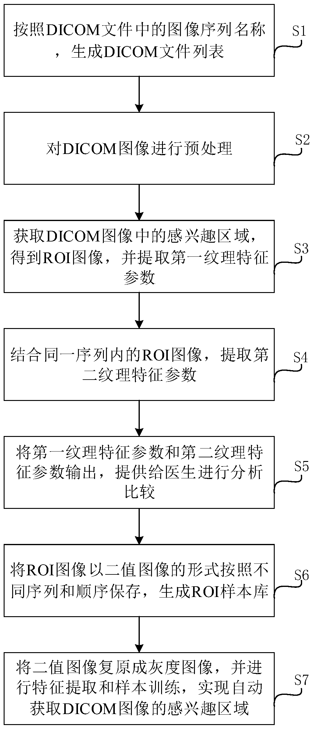
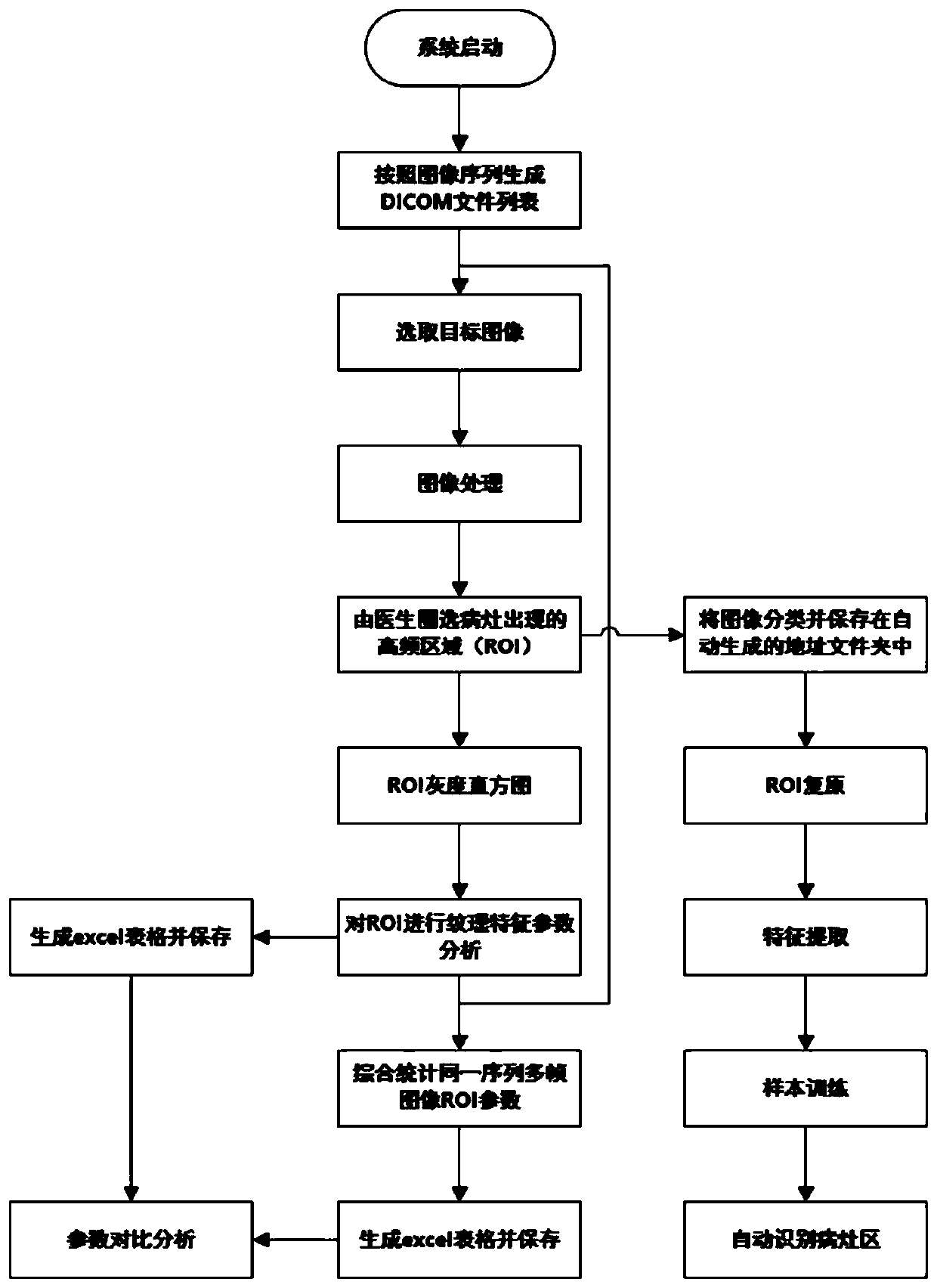
[0062] The present invention realizes the function of displaying and processing DICOM images on the PC, and the doctor can circle the high-frequency region that may appear in the lesion, that is, the region of interest (ROI region), and generate the ROI gray histogram. Perform texture analysis on the ROI area, and extract the first-order texture feature parameters (including histogram and mean, entropy, peak and skewness) and second-order texture feature parameters (including gray co-occurrence matrix, energy, entropy, inertial moments and correlations). And it can synthesize all ROI regions of the entire sequence of DICOM images, superimpose to generate a grayscale histogram, and extract the first-order texture feature parameters (histogram and mean, entropy, peak value, skewness) of the superimposed ROI region image. The sequence hierarchy identifies the type of modality that produced the image, the date the sequence was produced, details of the type of examination, equipmen...


PUM


Abstract

The invention relates to a DICOM image processing method which comprises the following steps: S1, generating a DICOM file list according to an image sequence name in a DICOM file; S2, preprocessing the DICOM image; S3, acquiring a region of interest in the DICOM image to obtain an ROI image, and extracting a first texture feature parameter; S4, in combination with the ROI images in the same sequence, extracting a second texture feature parameter; S5, outputting the first texture feature parameter and the second texture feature parameter. Compared with the prior art, the method has the advantages that the working efficiency and convenience of doctors are improved, and the like.

Description

technical field [0001] The invention relates to the field of image processing, in particular to a DICOM image processing method and system. Background technique [0002] Nowadays, medical images based on computerized tomography (Computed Tomography, CT) and magnetic resonance imaging (Magnetic Resonance Imaging, MRI) have the advantages of high tissue resolution, multi-sequence, and multi-directional imaging, and have been obtained in clinical examinations. It is widely used, but it is mostly recognized by doctors' manual experience. However, there are still many internal heterogeneities in benign and malignant tumors that are difficult to distinguish with the naked eye. For such cases, doctors often need to go through a complicated process to complete the pathological diagnosis. Experienced doctors cannot make quick and accurate judgments, which undoubtedly increases the burden on doctors and reduces their work efficiency. [0003] In machine vision and image processing, t...

Claims


Application Information

Patent Timeline
Patent Type & Authority Applications(China)
IPC IPC(8): G06K9/32G06T7/00G06T7/136G06T7/44G06T7/90G16H30/20G16H50/20
CPCG06T7/44G06T7/136G06T7/90G06T7/0012G16H30/20G16H50/20G06T2207/30004G06V10/25
Inventor 刘瑾赵潇杨海马李筠李芳付玏刘莹陈嘉慈
Owner SHANGHAI UNIV OF ENG SCI
Why Eureka
Try Eureka