

Medical image processing method and device, image processing equipment and storage medium
A medical image and processing method technology, applied in the field of medical image processing, can solve the problems of slow blood vessel segmentation and other problems
- Summary
- Abstract
- Description
- Claims
- Application Information
AI Technical Summary
Problems solved by technology
Method used
Image


Examples
Embodiment 1
[0027] figure 1 It is a flow chart of the medical image processing method provided by Embodiment 1 of the present invention. The technical solution of this embodiment is applicable to the case of improving the processing speed of the medical image by reducing the amount of edge padding of the target image. The method can be executed by the medical image processing apparatus provided in the embodiment of the present invention, and the apparatus can be implemented in software and / or hardware, and configured to be applied in a processor of a medical image processing device. The method specifically includes the following steps:
[0028] S101. Divide the target image into a plurality of analysis image blocks through a first sliding window, or jointly divide the target image into a plurality of analysis image blocks of corresponding sizes through at least two second sliding windows with different window edge sizes, Wherein, the size of the window edge in each direction of the firs...
Embodiment 2
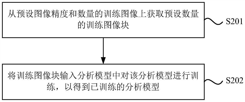
[0063] figure 2 It is a flow chart of the medical image processing method provided by Embodiment 2 of the present invention. On the basis of the foregoing embodiments, the embodiment of the present invention adds an explanation of the analysis model training method. Such as figure 2 As shown, the training method includes:
[0064] S201. Acquire a preset number of training image blocks from the preset image accuracy and number of training images.
[0065] Wherein, the preset image precision is preferably an image precision commonly used in clinical diagnostic images, and of course other image precisions, such as (1.0, 1.0, 1.0), may also be used. As long as the image precision of the training image blocks used to train the analysis model is the same as that of the analysis image blocks described in the foregoing embodiments.
[0066] The training images are clinical diagnostic images after image recognition processing. Taking a trained analysis model for analyzing CTA im...
Embodiment 3
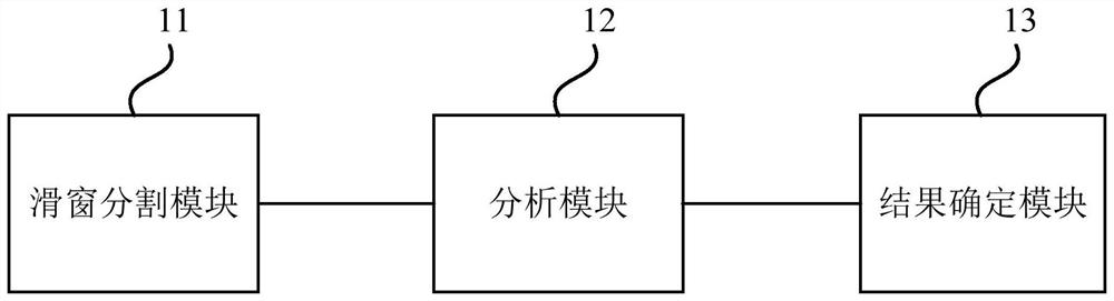
[0076] image 3 It is a structural block diagram of the medical image processing device provided by Embodiment 3 of the present invention. The device is used to execute the medical image processing method provided in any of the above embodiments, and the device may be implemented by software or hardware. The unit includes:
[0077] The sliding window segmentation module 11 is used to divide the target image into a plurality of analysis image blocks through the first sliding window, or jointly divide the target image into corresponding size blocks through at least two second sliding windows with different window edge sizes. A plurality of analysis image blocks, wherein, the size of the window edge in each direction of the first sliding window and the second sliding window is determined based on the principle of the minimum number of filling edges;
[0078] The analysis module 12 is used to input the analysis image blocks into the trained analysis model in batches to obtain th...
PUM
 Abstract
Description
Claims
Application Information