Medical perfusion image processing method and medical imaging equipment

An image processing and medical technology, applied in the field of image processing, can solve the problems of poor determination accuracy, time-consuming, and complicated determination process of arterial input points, and achieve the effect of avoiding human intervention and improving accuracy and efficiency

Pending Publication Date: 2020-10-30
SHANGHAI UNITED IMAGING HEALTHCARE
View PDF0 Cites 2 Cited by
  • Summary
  • Abstract
  • Description
  • Claims
  • Application Information

AI Technical Summary

Problems solved by technology

[0005] However, the arterial input point determination method in the prior art has the following problems: the manual selection method has high precision, but the determination process is complicated and time-consuming
The automatic selection method only considers the

Method used


Image

  • Medical perfusion image processing method and medical imaging equipment
  • Medical perfusion image processing method and medical imaging equipment
  • Medical perfusion image processing method and medical imaging equipment

Examples


Embodiment 1

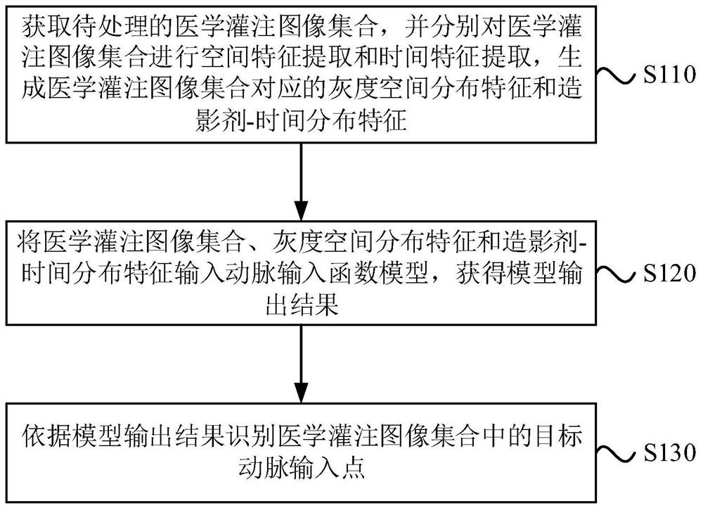
[0035] The medical perfusion image processing method provided in this embodiment is applicable to the situation of automatically identifying an arterial input point from a medical perfusion image. The method can be performed by a medical perfusion image processing device, which can be implemented by software and / or hardware, and the device can be integrated in electronic equipment with image processing functions, such as notebook computers, desktop computers, servers or magnetic resonance Image processing workstations of medical equipment such as imaging equipment and computer tomography imaging equipment, etc. see figure 1 , the method of this embodiment specifically includes:

[0036] S110. Acquire a medical perfusion image set to be processed, and perform spatial feature extraction and temporal feature extraction on the medical perfusion image set respectively, to generate grayscale spatial distribution features and contrast agent-time distribution features corresponding t...

Embodiment 2

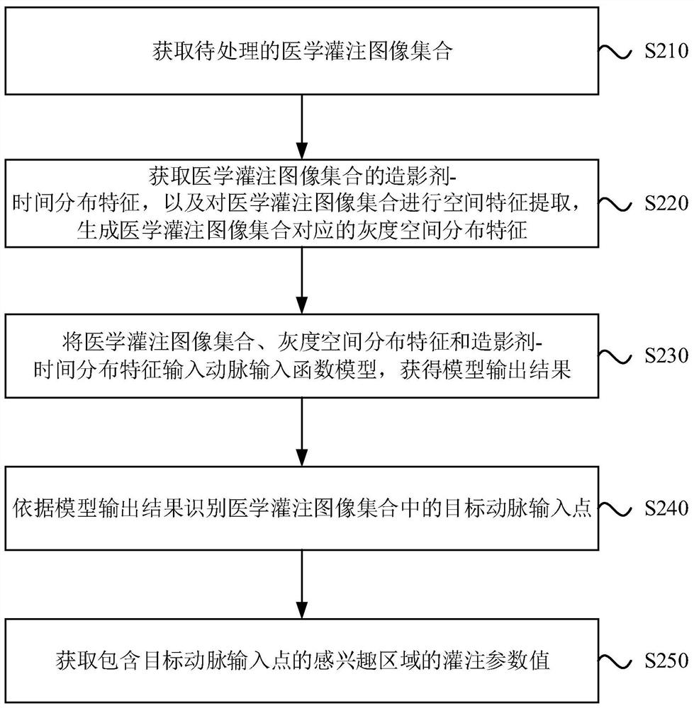
[0049] This embodiment provides a medical perfusion image processing method, which can be executed by a magnetic resonance system, a computed tomography system or a medical post-processing workstation, see image 3 , the specific steps of the method may include:

[0050] S210. Acquire a set of medical perfusion images to be processed.

[0051] The set of medical perfusion images includes images of one or more slices. The medical image to be processed can be obtained, for example, by hepatic blood perfusion imaging, for example, by performing dynamic scanning of the selected slice after intravenous bolus injection of contrast agent. Certainly, the region of interest corresponding to the medical image to be processed may also be organs or tissues such as the brain, heart, lungs, and kidneys.

[0052] A collection of medical perfusion images may be obtained by applying a bolus of contrast agent to a patient's vasculature, and imaging a region of interest at a number of differen...

Embodiment 3

[0069] In this embodiment, on the basis of the first embodiment above, the "arterial input function model" is further optimized. Wherein, explanations of terms that are the same as or corresponding to the above embodiments are not repeated here.

[0070] see Figure 6 , the model architecture of the arterial input function model in the medical perfusion image processing method provided in this embodiment includes: a recognition network unit 610 and a synthesis unit 620, and the recognition network unit 610 includes a space recognition sub-network 611 and a time recognition sub-network 612; wherein, the space The identification subnetwork 611 is used to identify the medical perfusion image set from the spatial dimension, the time identification subnetwork 612 is used to identify the medical perfusion image from the time dimension, and the synthesis unit 620 is used to obtain the model output result.

[0071] The spatial identification sub-network 611 is used to identify the ar...


PUM


Abstract

The embodiment of the invention discloses a medical perfusion image processing method and medical imaging equipment. The method comprises the steps: acquiring a medical perfusion image set to be processed, performing spatial feature extraction and time feature extraction on the medical perfusion image set, and generating gray scale spatial distribution features and contrast agent-time distributionfeatures corresponding to the medical perfusion image set; inputting the medical perfusion image set, the gray scale space distribution characteristics and the contrast agent-time distribution characteristics into an artery input function model to obtain a model output result; and identifying a target artery input point in the medical perfusion image set according to the model output result. According to the technical scheme, the artery input point can be recognized from the medical perfusion image more quickly and accurately.

Description

technical field [0001] Embodiments of the present invention relate to image processing technologies, and in particular to a medical perfusion image processing method and medical imaging equipment. Background technique [0002] Perfusion is a dynamic contrast-enhanced scanning examination method commonly used in modern medicine, and is currently mainly used in the clinical diagnosis of acute cerebral ischemic diseases or tumors. For cerebral ischemic diseases, perfusion imaging can reveal lesions as early as 30 minutes after ischemia. In addition, perfusion can also be used to assess the degree of ischemia, quantify and analyze hemodynamic changes in unit tissues, and provide reference for formulating correct thrombolytic methods. [0003] In the process of processing medical perfusion images, it is necessary to accurately obtain the position of the artery where the contrast agent first enters from the vein (that is, the arterial input point), and then obtain the contrast ag...

Claims


Application Information

Patent Timeline
IPC IPC(8): G06T11/00
CPCG06T11/003G06T2211/404G06T2211/424Y02A90/10
Inventor 王梅云宗金光
Owner SHANGHAI UNITED IMAGING HEALTHCARE
Why Eureka
Try Eureka