Artificial intelligence system for identifying high myopia retinopathy

A technology for retinopathy and high myopia, applied in the field of artificial intelligence systems, can solve problems such as low patient compliance, splitting, and complicated fundus performance in high myopia, and achieve the effect of saving inspection time

Pending Publication Date: 2020-12-08
ZHONGSHAN OPHTHALMIC CENT SUN YAT SEN UNIV
View PDF7 Cites 4 Cited by
  • Summary
  • Abstract
  • Description
  • Claims
  • Application Information

AI Technical Summary

Problems solved by technology

[0003] 1. The possibility of implementing large-scale crowd screening is low: At present, there are a large myopia population in my country, and medical resources are limited. There are 600 million myopia patients, and the overall myopia rate among teenagers is as high as 55%. Myopia patients account for about 21%. Such patients are prone to complications such as retinal atrophy, splitting, macular hole, retinal detachment, and choroidal neovascularization, which will cause irreversible damage to vision and seriously affect the living standards of young and middle-aged people.
[0004] 2. The fundus manifestations of high myopia are complex, and the requirements for inspectors are high: high myopia patients have severe retinal atrophy and many complications, and the clinical experience of the examiners must be high. Interpretation of high myopia retinopathy requires ophthalmologists to undergo professional training and comparison. With the accumulation of long-term experience, community hospitals or low-level general hospitals and physical examination centers usually do not have professional ophthalmologists, so they cannot conduct comprehensive detection and evaluation of such lesions.
[0005] 3. Patients with high myopia need life-long follow-up: Depending on the condition, patients with high myopia need to be followed up every 3-6 months. However, some remote areas and grassroots hospitals lack effective medical resources, and patients need to be followed up in general hospitals in other places. Repeated running will bring a greater burden to patients, which may easily lead to lower patient compliance and loss of follow-up
[0006] 4. OCT images (optical coherence tomography images) contain more information, but the images are more complex: Compared with other fundus imaging examinations such as fundus color photos, OCT can display more lesion information, but the images are more complex. It is complex and has higher requirements for doctors to read images. General practitioners do not have the ability to read OCT images, and specialists also need a long period of training to obtain a higher accuracy rate of image reading.
And currently there is no deep learning model specifically for OCT images on the market. In view of the complexity of OCT images for high myopia, it is necessary to train an efficient deep learning model for this situation, so as to quickly realize the screening of large-scale population

Method used


Image

  • Artificial intelligence system for identifying high myopia retinopathy

Examples


Embodiment

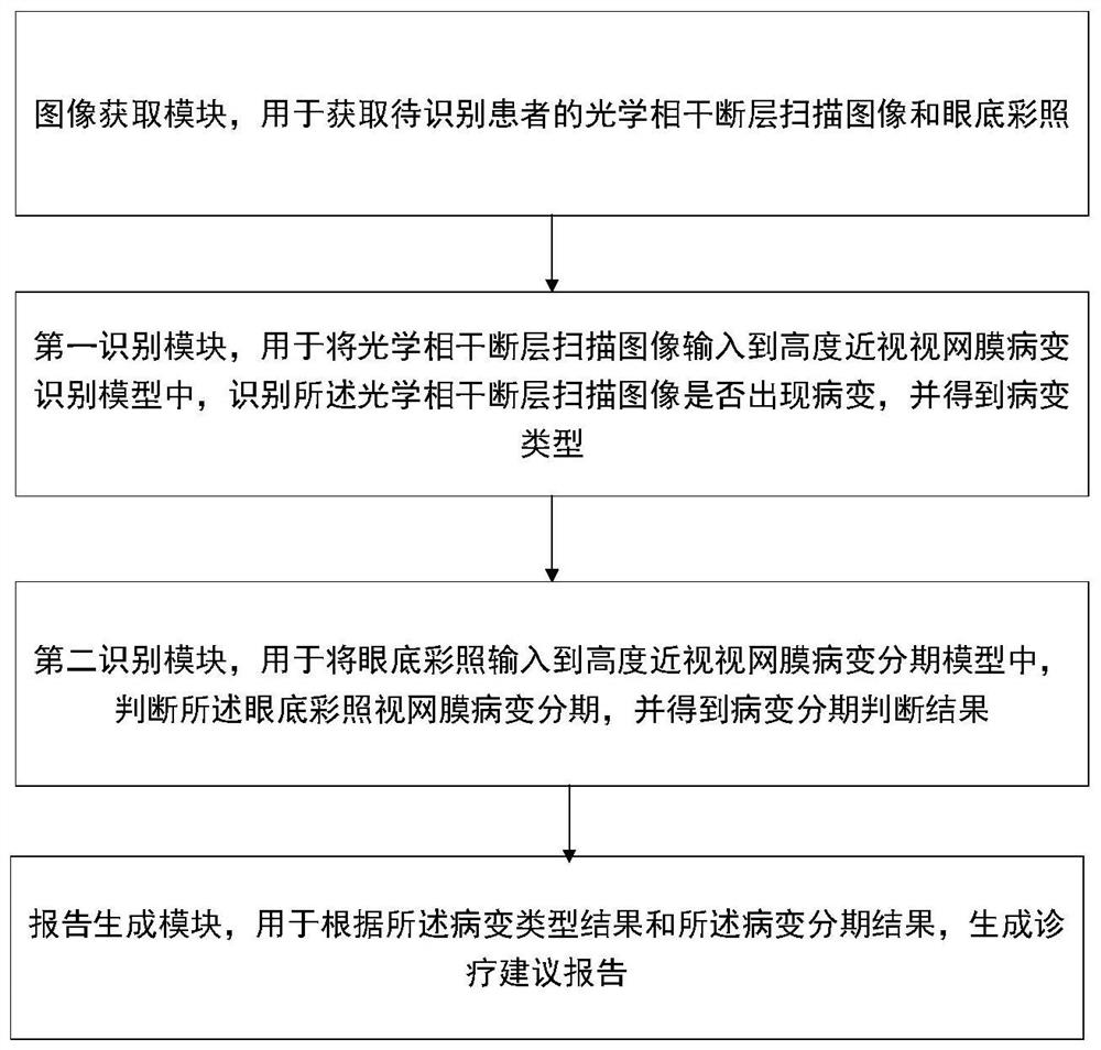
[0054] Such as figure 1 Shown is an artificial intelligence system for identifying high myopia retinopathy, the type of retinopathy includes retinoschisis, macular hole, retinal detachment, and choroidal neovascularization, and the system includes:

[0055] An image acquisition module, configured to acquire optical coherence tomography images and fundus color photos of patients to be identified;

[0056] The first identification module is used to input the optical coherence tomography image into the high myopia retinopathy identification model, identify whether there is a lesion in the optical coherence tomography image, and obtain the result of the lesion type;

[0057] Specifically, the training steps of the high myopia retinopathy recognition model include:

[0058] S1. Using multiple highly myopic retinal OCT image samples as a sample image set to classify according to retinoschisis, macular hole, retinal detachment, and choroidal neovascularization;

[0059] S2. Preproc...


PUM


Abstract

The invention relates to the technical field of medical image processing, in particular to an artificial intelligence system for identifying high myopia retinopathy, comprising: an image acquisition module for acquiring an optical coherence tomography image and a fundus reference of a patient to be identified; the first recognition module is used for inputting the optical coherence tomography image into a high myopia retinopathy recognition model, judging whether the optical coherence tomography image has lesion or not and obtaining a lesion type result; the second recognition module is used for inputting the fundus color photograph into a high myopia retinal lesion staging model, judging the retinal lesion staging of the fundus color photograph and obtaining a lesion staging judgment result; and the report generation module is used for generating a diagnosis and treatment suggestion report according to the lesion type result and the lesion staging judgment result. According to the method, the optical coherence tomography image and the fundus color photograph of the patient can be quickly and effectively recognized to recognize common retinopathy of high myopia.

Description

technical field [0001] The invention relates to the technical field of medical image processing, and more specifically, to an artificial intelligence system for identifying high myopia retinopathy. Background technique [0002] At present, the inspection process for high myopia retinopathy is as follows: first, the ophthalmologist performs refraction and axial measurement on the patient to determine whether the patient has high myopia and related risk factors, and then analyzes the patient's optical coherence tomography (OCT image) Determine whether there is retinopathy associated with high myopia (including retinoschisis, macular hole, retinal detachment, and choroidal neovascularization). It can be seen from the inspection process of high myopia retinopathy that it mainly has the following shortcomings: [0003] 1. The possibility of implementing large-scale crowd screening is low: At present, there are a large myopia population in my country, and medical resources are li...

Claims


Application Information

Patent Timeline
Patent Type & Authority Applications(China)
IPC IPC(8): G06T7/00G06K9/62G06N3/04G06N3/08A61B3/10A61B3/12A61B3/14
CPCG06T7/0012G06N3/08A61B3/102A61B3/12A61B3/14G06T2207/10004G06T2207/10016G06T2207/10101G06T2207/20081G06T2207/20084G06T2207/30041G06N3/045G06F18/241
Inventor 林浩添李永浩冯伟渤赵兰琴郭翀
Owner ZHONGSHAN OPHTHALMIC CENT SUN YAT SEN UNIV
Why Eureka
Try Eureka