Eye fundus image retinal vessel segmentation method based on mixed attention mechanism

A technology for retinal blood vessels and fundus images, applied in the field of image processing, can solve problems such as low precision, time-consuming pixel-level methods, and complicated operations, and achieve strong robustness

Active Publication Date: 2020-12-25
SHANTOU UNIV
View PDF6 Cites 17 Cited by
  • Summary
  • Abstract
  • Description
  • Claims
  • Application Information

AI Technical Summary

Problems solved by technology

When processing large-scale retinal images, pixel-level methods are time-consuming and difficult to meet clinical requirements
[0007] Generally speaking, the traditional image processing method is used to segment retinal blood vessels, which has high requirements on the image, and the operation is complicated and time-consuming. The segmentation effect obtained is not ideal and the accuracy is not high; It usual

Method used


Image

  • Eye fundus image retinal vessel segmentation method based on mixed attention mechanism
  • Eye fundus image retinal vessel segmentation method based on mixed attention mechanism
  • Eye fundus image retinal vessel segmentation method based on mixed attention mechanism

Examples


Embodiment 1

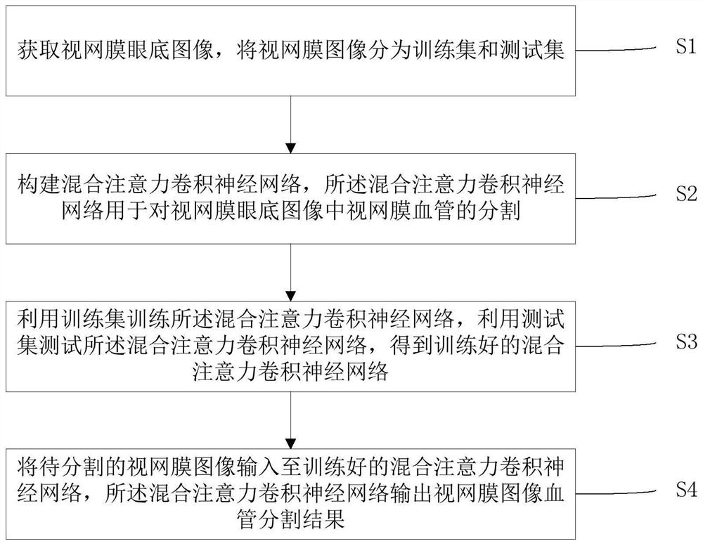
[0036] This embodiment provides a method for segmenting retinal blood vessels in fundus images with a mixed attention mechanism, such as figure 1 , including the following steps:

[0037] S1: Obtain the retinal fundus image, and divide the retinal image into a training set and a test set;

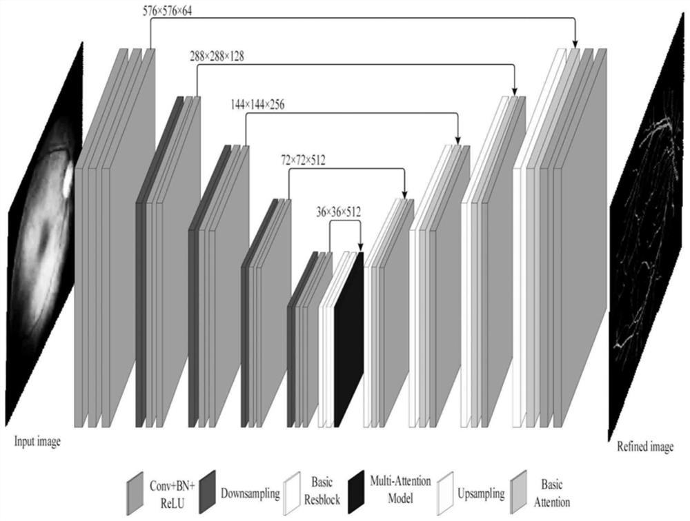
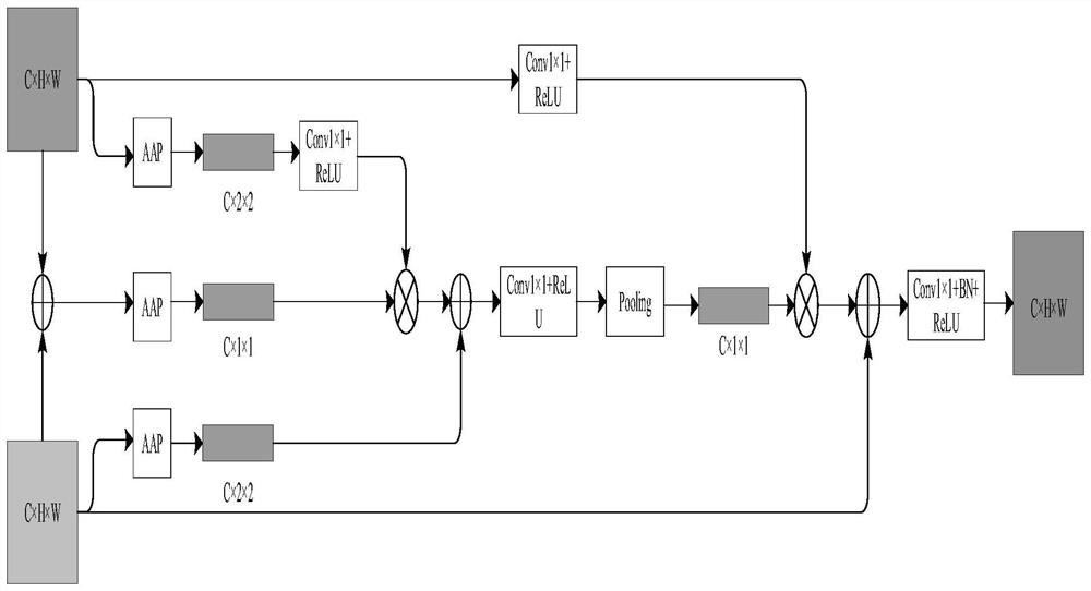
[0038] S2: Construct a mixed attention convolutional neural network, which is used to segment retinal blood vessels in retinal fundus images;

[0039] S3: using the training set to train the mixed attention convolutional neural network, and using the test set to test the mixed attention convolutional neural network to obtain a trained mixed attention convolutional neural network;

[0040]S4: Input the retinal image to be segmented into the trained mixed attention convolutional neural network, and the mixed attentional convolutional neural network outputs the retinal image blood vessel segmentation result, and the retinal image to be segmented can be obtained through the fundus stereo camer...


PUM


Abstract

The invention discloses an eye fundus image retinal vessel segmentation method based on a mixed attention mechanism, and the method comprises the following steps: S1, obtaining a retinal eye fundus image, and dividing the retinal image into a training set and a test set; S2, constructing a hybrid attention convolutional neural network, wherein the hybrid attention convolutional neural network is used for segmenting retinal vessels in the retinal fundus image; S3, training the hybrid attention convolutional neural network by using a training set, and testing the hybrid attention convolutional neural network by using a test set to obtain a trained hybrid attention convolutional neural network; and S4, inputting a to-be-segmented retinal image into the trained mixed attention convolutional neural network, wherein the mixed attention convolutional neural network outputs a retinal image blood vessel segmentation result. According to the method, a low-contrast vascular structure is effectively and accurately segmented, and the method has high robustness for interference of complex eye fundus image focuses, blood vessel center reflection phenomena and illumination imbalance phenomena.

Description

technical field [0001] The present invention relates to the field of image processing, and more specifically, to a method for segmenting retinal blood vessels in fundus images with a mixed attention mechanism. Background technique [0002] Retinal fundus image analysis is widely used in the diagnosis, screening and clinical research of eye diseases such as glaucoma and cataracts, cardiovascular diseases such as diabetes, hypertension and arteriosclerosis. Accurate segmentation of retinal vessels is the most important step in the analysis of retinal fundus images. Retinal vessels can not only reflect the condition of diseases such as diabetic retinopathy, but also help to locate and monitor retinal fundus lesions such as microaneurysms and hard exudates. diagnosis. However, in clinical practice, retinal vessel segmentation is generally completed by ophthalmologists or experts, which is a tedious and time-consuming task that requires skilled skills. Furthermore, different ob...

Claims


Application Information

Patent Timeline
IPC IPC(8): G06T7/00G06T7/10G06N3/04G06N3/08
CPCG06T7/0012G06T7/10G06N3/08G06T2207/20081G06T2207/20084G06T2207/30041G06T2207/30101G06N3/045
Inventor 马培立朱贵杰范衠李晓明林培涵
Owner SHANTOU UNIV
Why Eureka
Try Eureka