Cascaded cavity convolutional network brain tumor segmentation method with attention mechanism

A convolutional network and attention technology, applied in the field of 3D medical brain tumor image segmentation, can solve problems such as error-prone, time-consuming and labor-intensive, and achieve the effect of reducing the number imbalance, reducing the complexity, and reducing the consumption of network parameters and video memory

Inactive Publication Date: 2021-01-12
TIANJIN UNIV
View PDF11 Cites 7 Cited by
  • Summary
  • Abstract
  • Description
  • Claims
  • Application Information

AI Technical Summary

Problems solved by technology

However, manual segmentation by doctors is time-consuming and labor-intensive, and it is prone to errors after a long period of manual labeling. Doctors with different experience will have different segmentation results. Therefore, an automatic and accurate brain tumor segmentation method is needed.

Method used


Image

  • Cascaded cavity convolutional network brain tumor segmentation method with attention mechanism
  • Cascaded cavity convolutional network brain tumor segmentation method with attention mechanism

Examples


Embodiment Construction

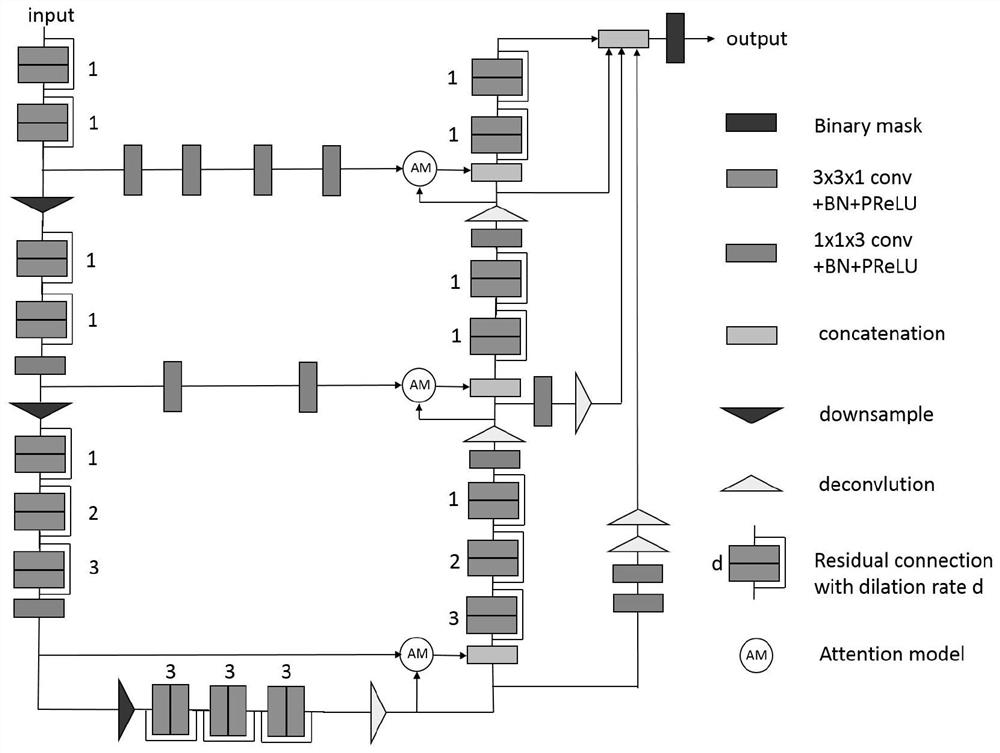
[0034] Firstly, the technical scheme of the brain tumor segmentation of the cascaded hollow convolutional network with attention mechanism of the present invention is introduced, and the steps are as follows:

[0035] (1) Data preprocessing:

[0036] This invention uses the published BraTS 2018 [6] The data set includes 285 cases in the training set and 66 cases in the verification set. The training set contains 210 cases of HGG and 75 cases of LGG. Each case has 3D MR images of four modes: T1, T1ce, T2 and FLAIR, and each size is 240×240×155; the validation set has 66 cases and does not distinguish between tumor types. In the axial, sagittal and coronal directions, it is divided into 144×144×19 blocks as the original input.

[0037] (2) Network structure construction, the method is as follows:

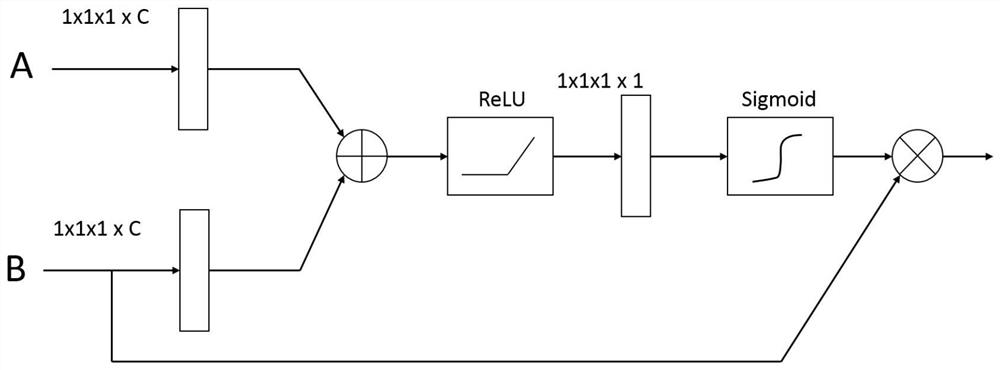
[0038] The cascaded hole convolutional network with attention mechanism adopts a three-level cascade framework, which simplifies multi-class segmentation tasks into three second-cl...


PUM


Abstract

The invention relates to a cascaded cavity convolutional network brain tumor segmentation method with an attention mechanism. The method comprises the following steps: data preprocessing; a network structure is built, and the method comprises the following steps that: a cascaded cavity convolutional network with an attention mechanism is built, a three-level cascaded framework is adopted, multipletypes of segmentation tasks are simplified into three second-class segmentation tasks, and the three-level segmented networks are respectively W-Net, T-Net and E-Net and are respectively used for segmenting a whole brain tumor (WT) region, a tumor nucleus (TC) region and an enhanced tumor nucleus (ET) region; segmentation is carried out from the axial direction, the sagittal direction and the coronal direction in each stage, then averaging is carried out in segmentation results in the three directions, and an accurate segmentation result is obtained; a network structure of each level in a three-level cascaded framework of cascaded hole convolution with an attention mechanism is a full convolutional network structure for encoding and decoding, and is divided into four parts, namely an encoder, a decoder, a skip layer structure and multi-layer feature map fusion.

Description

technical field [0001] The invention relates to the field of image processing, especially for three-dimensional medical brain tumor image segmentation. Background technique [0002] Brain tumors are intracranial tumors with high lethality. According to histological heterogeneity and tumor aggressiveness, brain tumors can be divided into high glial tumors (HGG) and low glial tumors (LGG). Brain tumors It can be further divided into edema area, tumor nuclear area, enhancing tumor nuclear area, non-enhancing tumor nuclear area, and necrotic area. The four modality images of brain tumor magnetic resonance (MR): T1, T1ce, T2 and FLAIR focus on different tumor areas and can provide complementary information for each other. Brain tumor segmentation is to separate different tumor regions in brain images, which is very important for patient disease assessment, treatment plan formulation and follow-up observation research. However, it is time-consuming and labor-intensive to rely on...

Claims


Application Information

Patent Timeline
Patent Type & Authority Applications(China)
IPC IPC(8): G06T7/11G06K9/62G06N3/04G06N3/08
CPCG06T7/11G06N3/08G06T2207/10072G06T2207/30096G06N3/045G06F18/253
Inventor 褚晶辉黄凯隆吕卫
Owner TIANJIN UNIV
Why Eureka
Try Eureka