Lateral chest radiography bone suppression method based on deep convolutional neural network

A convolutional neural network and neural network technology, which is applied in the field of lateral chest radiograph bone suppression based on deep convolutional neural network, can solve the problems of difficulty in decomposing lateral chest radiographs, overlapping and interlaced bones, and low contrast. Lung disease missed detection rate, improved accuracy, and the effect of avoiding occlusion

Active Publication Date: 2021-02-19
SOUTHERN MEDICAL UNIVERSITY
View PDF4 Cites 0 Cited by
  • Summary
  • Abstract
  • Description
  • Claims
  • Application Information

AI Technical Summary

Problems solved by technology

The lateral chest radiograph can provide valuable information and is often used as a supplementary image to the anterior-posterior chest radiograph, but the lateral chest radiograph has a deeper longitudinal length than the anteroposterior chest radiograph, has complex components, overlapping bones, and low contrast. Anterior-posterior chest radiographs, lateral chest radiographs are more difficult to decompose

Method used


Image

  • Lateral chest radiography bone suppression method based on deep convolutional neural network
  • Lateral chest radiography bone suppression method based on deep convolutional neural network
  • Lateral chest radiography bone suppression method based on deep convolutional neural network

Examples


Embodiment 1

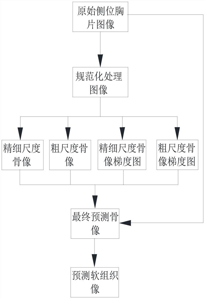
[0050] A bone suppression method for lateral chest radiographs based on deep convolutional neural networks, such as Figures 1 to 5 As shown, the steps involved are:

[0051] Step 1, performing normalization processing on the gray space of the original lateral chest radiograph image to obtain a normalized processing image;

[0052] Perform differential operations on the horizontal and vertical directions of the original lateral chest image to obtain a lateral chest gradient image, and then normalize the gray-scale space of the lateral chest gradient image to obtain a normalized processed gradient image;

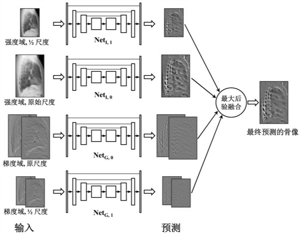
[0053] Step 2. Input the normalized processed image into the constructed fine-scale bone-suppressed convolutional neural network model in the intensity domain to obtain a fine-scale bone image;

[0054] Downsample the normalized image to the original size by 2 -s times to obtain a downsampled image, and then input the downsampled image into the coarse-scale bone-suppressed ...

Embodiment 2

[0088] A bone suppression method for lateral thoracic radiographs based on a deep convolutional neural network, specifically as follows:

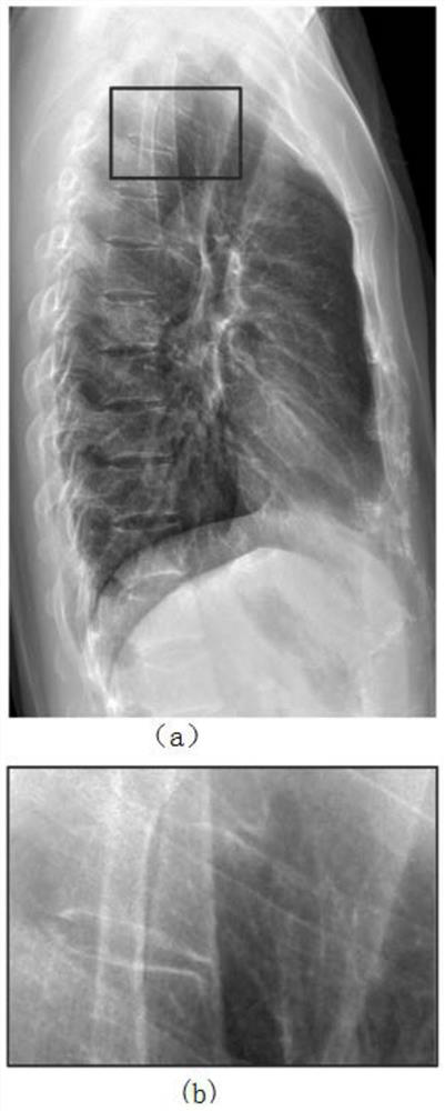
[0089] Step 1. For the input original lateral chest image I 0 , using a Gaussian image filter with a large filter kernel to image I 0 Perform smoothing and filtering to get the base map I b , from image I 0 Subtract Basemap I b , to get the detail map I d . In order to make the input of the bone suppression model consistent, the details of Figure I d The gray value of the normalized processing, so that I d The mean value of the gray level in is 0 and the variance is 1. Detail map I after grayscale normalization d Input for the fine-scale bone suppression model in the intensity domain.

[0090] Step 2. Use the fine-scale bone-suppressed convolutional neural network model in the intensity domain, and use the normalized lateral chest image obtained in step 1 as input to predict the bone image of the lateral chest image I, and multiply...


PUM


Abstract

The invention discloses a lateral chest radiography bone suppression method based on a deep convolutional neural network. A predicted soft tissue image is obtained through four steps. According to thelateral chest radiography bone suppression method based on the deep convolutional neural network, bone components and soft tissue components in a lateral chest radiography image can be separated, andthe soft tissue components are prevented from being shielded by the bone components in the lateral chest radiography. A multi-scale convolutional neural network model is trained by using dual-energysubtraction data in an intensity domain and a gradient domain, and a bone image and a soft tissue image which are finally predicted are obtained by fusing prediction results obtained by inputting a given lateral chest radiograph into each model under the maximum posterior probability, so that the bone image and the soft tissue image are obtained from a single lateral chest radiograph image. According to the method, bone components in the lateral chest radiography can be inhibited to serve as image supplementary information of the orthotopic chest radiography, so that the radiography reading accuracy is improved, and the omission ratio of lung diseases is reduced.

Description

technical field [0001] The invention relates to the technical field of medical image processing, in particular to a bone suppression method for lateral thoracic radiographs based on a deep convolutional neural network. Background technique [0002] Chest X-rays contain important lung information. However, due to the soft tissue shielding by bone components in chest X-rays, it is difficult for radiologists to read the films, resulting in a high rate of missed detection of pulmonary nodules. Although the soft tissue images obtained by dual-energy subtraction equipment can reduce the difficulty of film reading and reduce the missed detection rate of pulmonary nodules, due to the fact that dual-energy subtraction equipment has not yet been popularized in major hospitals and the radiation dose is high, it is not the most The preferred imaging examination. The X-ray chest film is the most preferred technical method at present. Based on image processing technology, the conventiona...

Claims


Application Information

Patent Timeline
Patent Type & Authority Applications(China)
IPC IPC(8): G06T7/00G06T7/90G06T5/20G06N3/08G06N3/04G06K9/62
CPCG06T7/0012G06T5/20G06T7/90G06N3/08G06T2207/20084G06T2207/20081G06T2207/30008G06T2207/10116G06N3/045G06F18/214
Inventor 阳维刘云碧席誉华秦耿耿冯前进
Owner SOUTHERN MEDICAL UNIVERSITY
Why Eureka
Try Eureka