
Cardiac ejection fraction calculation method and device

A technology of cardiac ejection and calculation method, applied in the field of video processing, can solve the problems of error, complicated operation, subjectivity and unreliability of calculation results, etc., and achieve the effect of high accuracy and strong ease of use

Pending Publication Date: 2021-02-19
深圳蓝影医学科技股份有限公司
View PDF0 Cites 1 Cited by
  • Summary
  • Abstract
  • Description
  • Claims
  • Application Information

AI Technical Summary

Problems solved by technology

[0004] However, due to technical limitations, the three-dimensional echocardiography method is seldom used clinically and is mostly used in the research stage; the M-mode echocardiography method (M-mode echocardiography method uses a single sound beam to scan the heart, because the display Cardiovascular movement, so it is named M-mode echocardiography according to the first letter "M" of English movement) The left ventricular ejection fraction obtained by two-dimensional echocardiography is less accurate; the left ventricular ejection fraction obtained by two-dimensional echocardiography Although the ejection fraction has high accuracy, the operation is cumbersome, especially the manual endocardium drawing, which is not only time-consuming and labor-intensive but also prone to operator errors, and the calculation results are subject to greater subjectivity and unreliability

Method used


Image

  • Cardiac ejection fraction calculation method and device
  • Cardiac ejection fraction calculation method and device
  • Cardiac ejection fraction calculation method and device

Examples


Embodiment Construction

[0030] In order to make the purpose, features and advantages of the present application more obvious and understandable, the present application will be further described in detail below in conjunction with the accompanying drawings and specific implementation methods. Apparently, the described embodiments are some of the embodiments of the present application, but not all of them. Based on the embodiments in this application, all other embodiments obtained by persons of ordinary skill in the art without creative efforts fall within the protection scope of this application.

[0031] It should be noted that, in any embodiment of the present invention, a method for calculating cardiac ejection fraction is used to calculate cardiac ejection fraction by using ultrasound data of a target heart region.

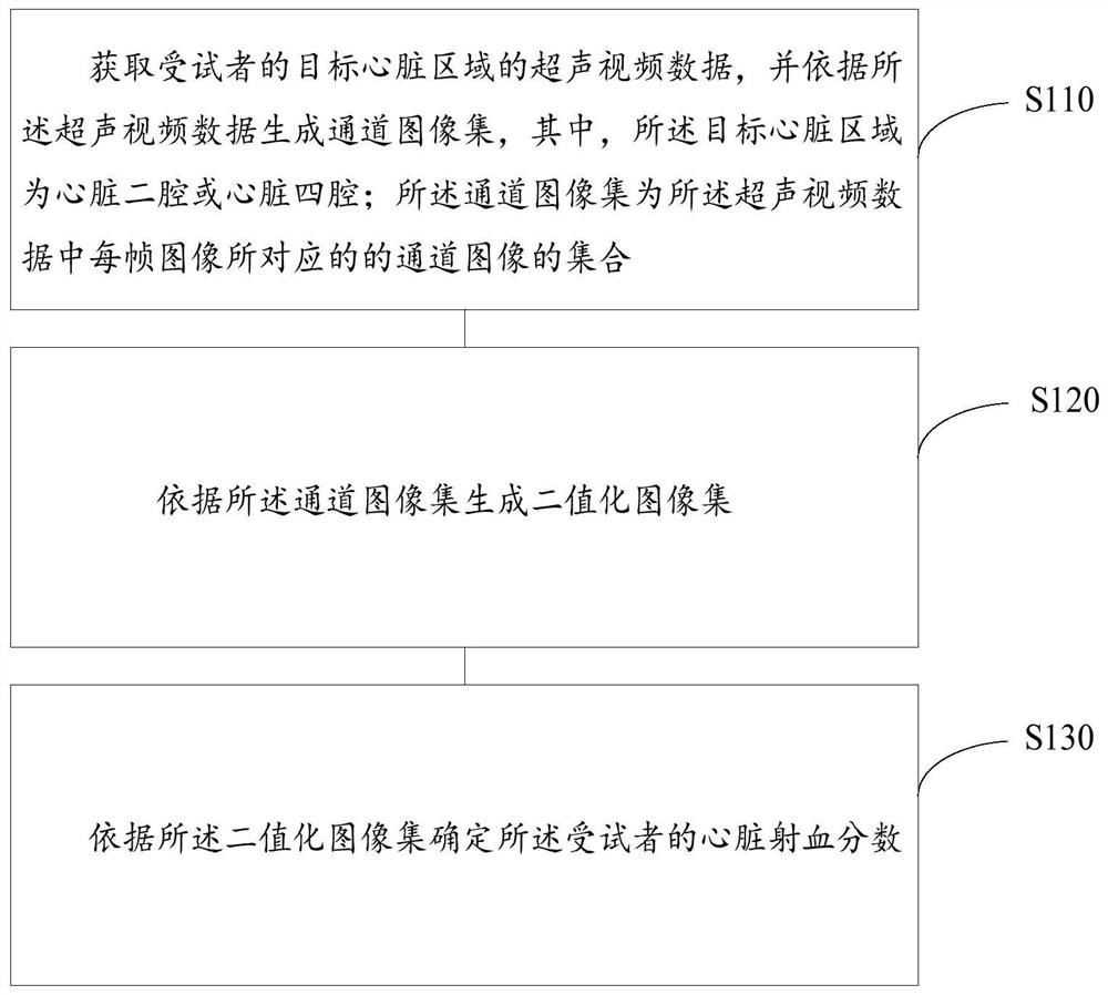
[0032] refer to figure 1 , which shows a flow chart of the steps of a method for calculating cardiac ejection fraction provided by an embodiment of the present application, specifi...


PUM


Abstract

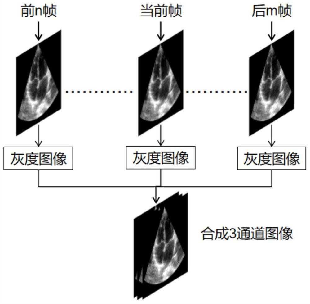
The embodiment of the invention provides a cardiac ejection fraction calculation method and device, and the method comprises the steps: obtaining ultrasonic video data of a target cardiac region of asubject, and generating a channel image set according to the ultrasonic video data, wherein the target heart region is a cardiac two-cavity or heart four-cavity; wherein the channel image set is a setof channel images corresponding to each frame of image in the ultrasonic video data; generating a binary image set according to the channel image set; and determining the cardiac ejection fraction ofthe subject according to the binarized image set. By utilizing the self-learning function of the neural network, the cardiac ejection fractions of multiple cardiac cycles are automatically calculated, the defects of a traditional calculation method are overcome, the method does not depend on other devices such as electrocardiogram devices and cross-cardiac cycle analysis devices, and the invention has the advantages of being high in accuracy, high in usability, repeatable and the like. Sufficient and accurate reference data is provided for classification, diagnosis and treatment of heart failure.

Description

technical field [0001] The invention relates to the technical field of video processing, in particular to a method and device for calculating cardiac ejection fraction. Background technique [0002] Cardiac ejection fraction is an important clinical indicator for evaluating cardiac function and a reference for the classification, diagnosis and treatment of heart failure. Cardiac ejection fraction refers to the ratio of the blood output produced by the heart per beat to the end-diastolic volume of the left ventricle. [0003] Calculation methods of cardiac ejection fraction mainly include echocardiography, two-dimensional echocardiography and three-dimensional echocardiography, wherein the two-dimensional echocardiography is also called the modified biplane Simpson method. [0004] However, due to technical limitations, the three-dimensional echocardiography method is seldom used clinically and is mostly used in the research stage; the M-mode echocardiography method (M-mode ...

Claims


Application Information

Patent Timeline
IPC IPC(8): G06T7/90G06T7/62G06T7/11G06N3/08G06N3/04
CPCG06T7/11G06T7/62G06T7/90G06N3/08G06T2207/10136G06T2207/30048G06N3/045
Inventor 杨勇邢锐桐陈晶丁浩
Owner 深圳蓝影医学科技股份有限公司
Learn More