

Lung gland squamous cell carcinoma diagnosis device based on PET/CT image sub-region imaging omics characteristics
A radiomics and diagnostic device technology, applied in the field of medical imaging and machine learning, can solve the problem of low diagnostic efficiency and achieve the effect of improving accuracy
- Summary
- Abstract
- Description
- Claims
- Application Information
AI Technical Summary
Problems solved by technology
Method used
Image

Examples
Embodiment Construction
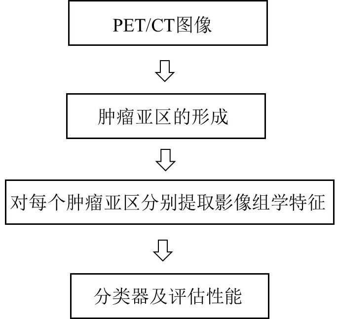
[0026] The method of the present invention is obvious in clinical imaging according to the regional changes in the tumor. Specifically, in a single tumor, the intratumoral surface is different, such as necrotic area and highly active area, which reflect different biological processes. Therefore, the tumor is first subjected to sub-regional partition extraction processing ( figure 2 ), and then perform radiomics feature extraction respectively, so as to better consider tumor heterogeneity, extract more effective radiomics features, and improve the accuracy of tumor diagnosis. Specifically, a lung squamous adenocarcinoma diagnostic device based on radiomics features of PET / CT image subregions of the present invention, the diagnostic device specifically includes:
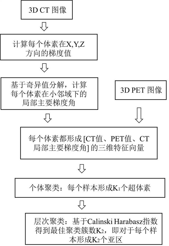
[0027] A voxel three-dimensional feature extraction module, the voxel three-dimensional feature extraction module is used to extract the CT local main gradient angle feature value of each voxel of the lung tumor in a ...
PUM
 Abstract
Description
Claims
Application Information