
Cell drug resistance detection method based on high content imaging, medium and electronic equipment

A detection method and drug resistance technology are applied in the field of image processing and automatic recognition of convolutional neural networks, which can solve the problems of heavy workload of pathological reading and achieve the effect of improving recognition accuracy.

Pending Publication Date: 2021-03-16
SHANGHAI UNIV OF MEDICINE & HEALTH SCI +1
View PDF0 Cites 2 Cited by
  • Summary
  • Abstract
  • Description
  • Claims
  • Application Information

AI Technical Summary

Problems solved by technology

In clinical practice, doctors will make relevant diagnoses based on cell pictures, but the workload of pathological film reading is huge

Method used


Image

  • Cell drug resistance detection method based on high content imaging, medium and electronic equipment
  • Cell drug resistance detection method based on high content imaging, medium and electronic equipment
  • Cell drug resistance detection method based on high content imaging, medium and electronic equipment

Examples


Embodiment 1

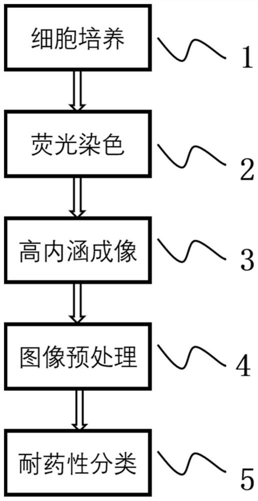
[0033] This embodiment provides a method for detecting drug resistance of cells based on high-content imaging, such as figure 1 shown, including the following steps:

[0034] Step 1, culturing target cells and drug-resistant strains thereof. In this example, 24-well glass plates were used, and 50,000 cells per well were cultured for 36 hours to allow the cells to adhere to the wall. The culture in this example involves PC9 and PC9 / GR cells.

[0035] Step 2, select a suitable fluorescent dye for the selected cells to stain.

[0036] In this example, the cultured cells were stained in the following manner:

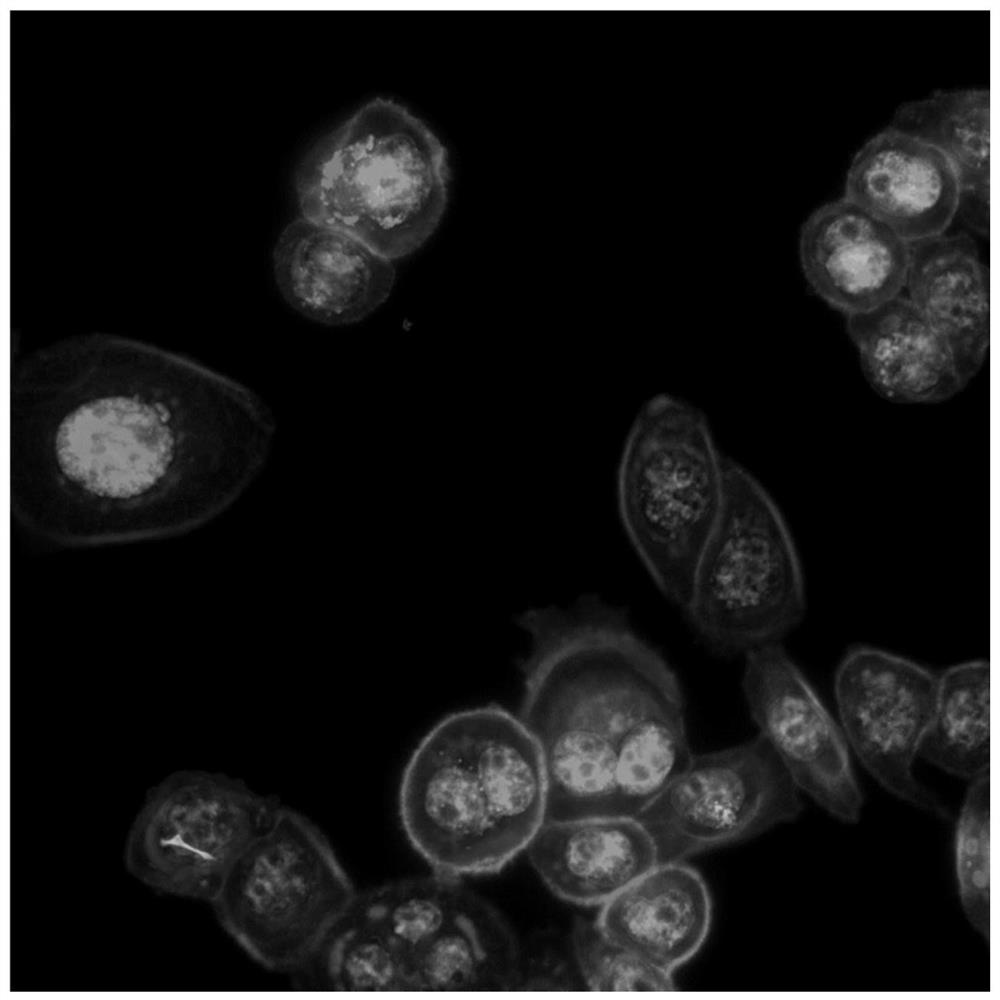
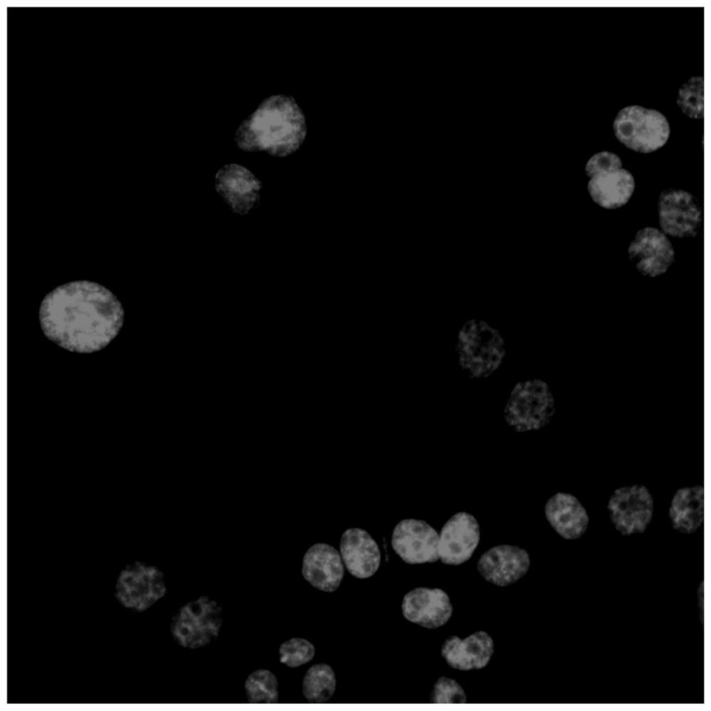
[0037] (21) Discard the culture medium, wash twice with 2 ml of PBS, take Mito Tracker Deep Red FM stock solution (1mM with DMSO) and add PBS to dilute to 250nM, add to each well, put in the incubator and continue to incubate for 20 minutes;

[0038] (22) Wash 3 times with PBS, 10 minutes each time, suck away the cleaning solution, fix the cells with 4% paraformaldehyde f...

Embodiment 2

[0069] This embodiment provides an electronic device, including one or more processors, a memory, and one or more programs stored in the memory, the one or more programs including the high-level Instructions for endogenous imaging methods for the detection of cellular drug resistance.


PUM


Abstract

The invention relates to a cell drug resistance detection method based on high content imaging, a medium and electronic equipment. The cell drug resistance detection method comprises the following steps: carrying out fluorescence staining on cells to be detected; performing high-content imaging on the dyed to-be-detected cells to obtain a corresponding high-content image; preprocessing the high content image; and taking the preprocessed image as the input of a trained drug resistance classification and identification model based on the convolutional neural network, and detecting the drug resistance category to which the to-be-detected cell belongs. Compared with the prior art, the method has the advantages of simplicity, reliability, high efficiency and the like.

Description

technical field [0001] The invention belongs to the field of image processing and convolutional neural network automatic identification, and in particular relates to a cell drug resistance detection method, medium and electronic equipment based on high-content imaging. Background technique [0002] The identification of drug resistance of research cells can not only help researchers to effectively identify and screen drug-resistant cell lines in the laboratory, but also help doctors to accurately judge the drug resistance of patients clinically. At present, the identification of drug resistance of cancer cells at home and abroad is mainly based on the current method of laboratory identification of drug resistance of cell lines, which mainly uses flow cytometry, MTT method and gene chip for identification. Clinical judgment of drug resistance is mainly based on drug sensitivity test or drug resistance gene detection. These methods are difficult and difficult to guarantee eff...

Claims


Application Information

Patent Timeline
IPC IPC(8): G06K9/00G06K9/62G06N3/04G01N21/64G01N15/14
CPCG01N21/6458G01N15/1434G01N2015/144G06V20/698G06N3/047G06N3/045G06F18/2415
Inventor 黄钢李秀英聂生东
Owner SHANGHAI UNIV OF MEDICINE & HEALTH SCI
Learn More