Automatic segmentation method for various tissues in mouse testis pathological section based on deep learning

A technology of pathological slices and deep learning, applied in neural learning methods, image analysis, image data processing, etc.

Pending Publication Date: 2021-03-19
NANJING UNIV OF INFORMATION SCI & TECH
View PDF0 Cites 0 Cited by
  • Summary
  • Abstract
  • Description
  • Claims
  • Application Information

AI Technical Summary

Problems solved by technology

Automatic segmentation of multiple types of germ cells and multiple types of tissue regions is a prerequisite for establishing an automatic staging...

Method used


Image

  • Automatic segmentation method for various tissues in mouse testis pathological section based on deep learning
  • Automatic segmentation method for various tissues in mouse testis pathological section based on deep learning
  • Automatic segmentation method for various tissues in mouse testis pathological section based on deep learning

Examples


specific Embodiment

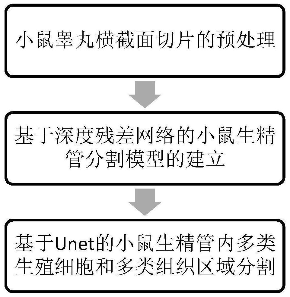
[0052] 1. First, color-label all the pathological images of the mouse testis cross-section;

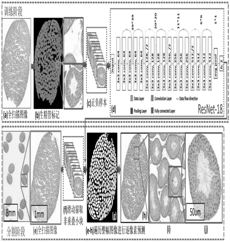
[0053] 2. Then, scale a full scan image of a mouse testis cross section to 1 / 400 of the original size (the length and width are reduced by 20 times), and then send it to the deep convolutional neural network for pixel-by-pixel segmentation to obtain the mouse Pre-segmentation results of seminiferous tubules. Use bilinear interpolation to map the segmentation result to the size of the original image;

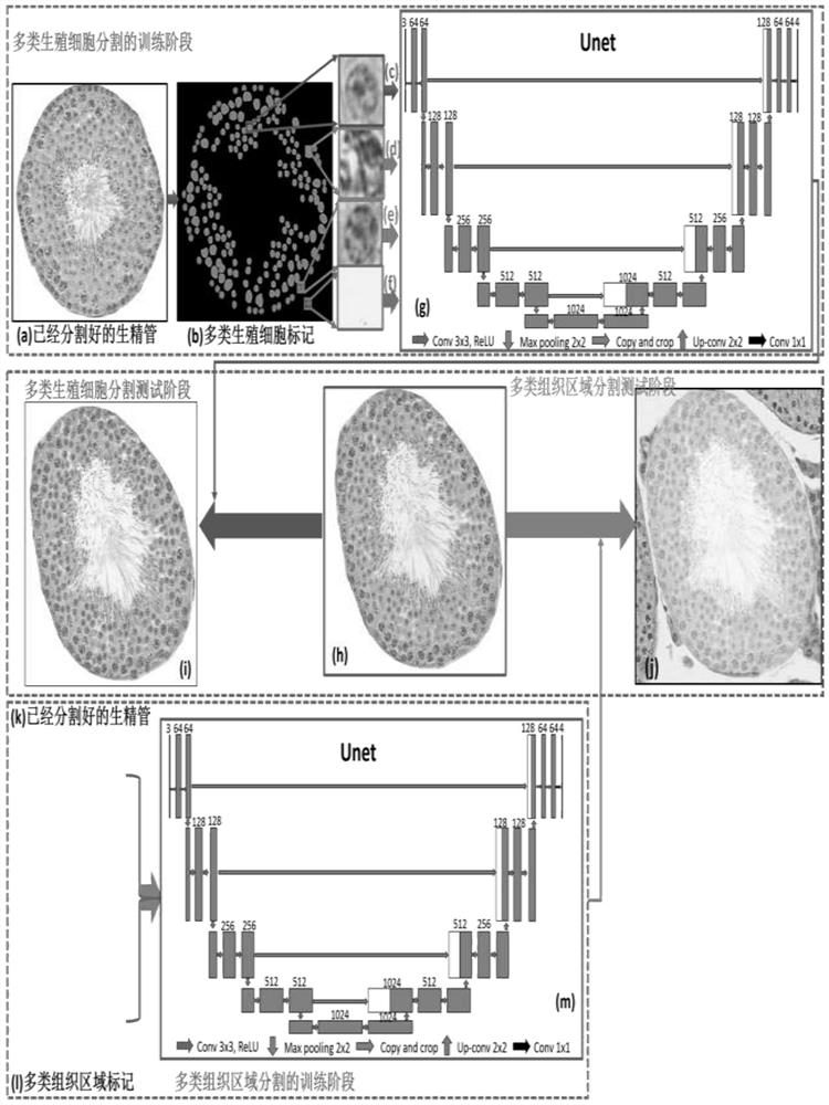
[0054] 3. In the staging of seminiferous ducts in mice, stages VII-VIII are the two consecutive stages that are most difficult for pathologists to distinguish, so it is planned to initially analyze the images of stages VII-VIII that are most difficult to distinguish; The seminiferous tubules sorted out in the first stage were extracted, and Unet was used to perform multi-type germ cell segmentation and multi-type tissue region segmentation.


PUM


Abstract

The invention discloses an automatic segmentation method for various tissues in a mouse testis pathological section based on deep learning. The method belongs to the field of machine learning and image processing. The method comprises the following specific steps: pretreating mouse testis cross section slices; establishing a mouse seminiferous tubule segmentation model based on ResNet; and establishing Unet-based segmentation of multiple types of germ cells and multiple types of tissue regions in the mouse seminiferous tubule. Firstly, seminiferous tubule pre-segmentation performedon a mouse testis cross section full-scanning image by using ResNet in combination with a sliding window and a pixel-by-pixel segmentation method; then, multiple types of cell nucleuses and multiple types of tissue regions in the seminiferous tubule are respectively segmented by using Unet. According to the method, good performance is obtained, and a good image analysis basis is provided for establishing a mouse seminiferous tubule automatic staging system; in the future, technicians extract histological characteristics of cell nucleuses and tissue regions in the mouse seminiferous tubule, and the histological characteristics are used for training an automatic staging classifier of the mouse seminiferous tubule.

Description

technical field [0001] The present invention relates to the fields of machine learning and image processing, in particular to an automatic segmentation method for various tissues in mouse testicular pathological sections based on deep learning. Background technique [0002] In an attempt to understand infertility caused by pathological defects in the human testis, early studies of the lesion were often modeled in mammals such as mice because the mammalian spermatogenesis process is relatively similar. At present, pathologists mainly rely on manual analysis for the staging of seminiferous ducts in mouse testes. Staging is based on image-based biomarkers such as the shape, texture, and distribution of various germ cells during spermatogenesis. This work is very important. Time-consuming, and the method is still relatively complicated and difficult to accurately identify the stage. Furthermore, since the pathologist's experience often determines his staging judgment, manual st...

Claims


Application Information

Patent Timeline
IPC IPC(8): G06T7/11G06T7/00G06T5/00G06N3/04G06N3/08
CPCG06T7/11G06T7/0012G06T5/007G06N3/08G06T2207/20081G06T2207/20084G06T2207/30004G06N3/045
Inventor 鲁浩达徐军闫朝阳
Owner NANJING UNIV OF INFORMATION SCI & TECH
Why Eureka
Try Eureka