Method for establishing angiography enhanced three-dimensional stenosis analysis model

A three-dimensional model and angiography technology, applied in the field of image processing, can solve the problems of analyzing data that cannot be visualized for the degree of vascular stenosis, is not conducive to the positioning and analysis of vascular lesion areas, and obtains real information of blood vessels, so as to achieve comprehensive blood vessel information and improve registration Efficiency and the effect of improving registration accuracy

Inactive Publication Date: 2021-04-09
XIAN CREATION KEJI CO LTD
View PDF0 Cites 1 Cited by
  • Summary
  • Abstract
  • Description
  • Claims
  • Application Information

AI Technical Summary

Problems solved by technology

However, the two-dimensional image has limitations, which is not conducive to obtaining the real information of blood vessels easily and quickly.
Moreover, from the images obtained by the above-mentioned imaging methods, it is impossible to intuitively and quickly obtain analysis data on the degree of vascular stenosis, which is not conducive to the clinical positioning and analysis of vascular lesion regions.

Method used


Image

  • Method for establishing angiography enhanced three-dimensional stenosis analysis model
  • Method for establishing angiography enhanced three-dimensional stenosis analysis model
  • Method for establishing angiography enhanced three-dimensional stenosis analysis model

Examples


Embodiment Construction

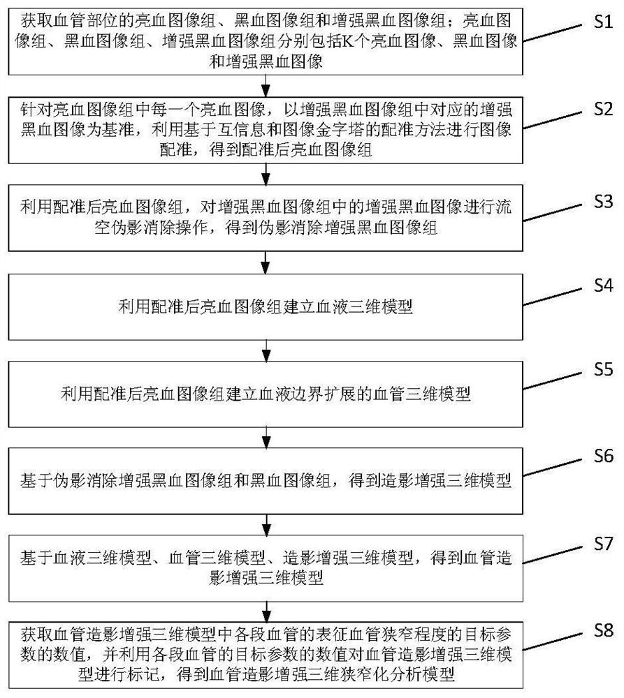
[0033] The technical solutions in the embodiments of the present invention will be described below with reference to the drawings in the embodiments of the present invention.

[0034] In order to obtain the real information of blood vessels and the analysis data about the degree of stenosis of blood vessels in a simple, fast and intuitive way in clinical application, so as to analyze vascular lesions. An embodiment of the present invention provides a method for establishing an angiography-enhanced three-dimensional stenosis analysis model.

[0035] It should be noted that the implementation subject of the method for establishing an angiography-enhanced three-dimensional stenosis analysis model provided by the embodiment of the present invention may be a device for establishing an angiography-enhanced three-dimensional stenosis analysis model, which can run on an electronic in the device. Wherein, the electronic device may be a blood vessel imaging device, or an image processi...


PUM


Abstract

The invention discloses a method for establishing an angiography enhanced three-dimensional stenosis analysis model. The method comprises the following steps: acquiring a bright blood image group, a black blood image group and an enhanced black blood image group of a blood vessel part; performing image registration on each bright blood image by using a registration method based on mutual information and an image pyramid to obtain a registered bright blood image group; carrying out flow empty artifact elimination operation on the enhanced black blood image by utilizing the registered bright blood image group to obtain an artifact-eliminated enhanced black blood image group; establishing a blood three-dimensional model and a blood vessel three-dimensional model by using the registered bright blood image group; obtaining a contrast enhanced three-dimensional model based on the artifact elimination enhanced black blood image group and the black blood image group; obtaining an angiography enhanced three-dimensional model; obtaining values of target parameters representing the vascular stenosis degree of each section of blood vessel in the angiography enhanced three-dimensional model, and marking the angiography enhanced three-dimensional model by utilizing the values of the target parameters of each section of blood vessel to obtain the angiography enhanced three-dimensional stenosis analysis model.

Description

technical field [0001] The invention belongs to the field of image processing, and in particular relates to a method for establishing an angiography-enhanced three-dimensional stenosis analysis model. Background technique [0002] According to the latest medical data, vascular diseases have seriously affected the life and health of contemporary people and become one of the diseases with a high mortality rate. Such as atherosclerosis, inflammatory vascular disease, vascular true neoplastic disease and so on. The common causes of vascular diseases are narrowing, blockage, rupture, and plaque in blood vessels. At present, methods based on lumen imaging are usually used to evaluate the degree of vascular disease and vascular stenosis clinically, such as digital subtraction angiography (Digital Subtraction Angiography, DSA), CT angiography (Computed Tomography Angiography, CTA), magnetic Resonance angiography (Magnetic Resonance Angiography, MRA) and high-resolution magnetic re...

Claims


Application Information

Patent Timeline
Patent Type & Authority Applications(China)
IPC IPC(8): G06T11/00G06T7/00G06T7/13G06T7/136G06T7/33G06T5/30G06T17/00G16H30/20G16H30/40
CPCG06T5/30G06T7/0012G06T11/008G06T17/00G06T2207/10088G06T2207/20016G06T2207/20024G06T2207/30096G06T2207/30101G06T2211/424G06T7/13G06T7/136G06T7/344G16H30/20G16H30/40
Inventor 贾艳楠王文杰
Owner XIAN CREATION KEJI CO LTD
Why Eureka
Try Eureka