Cell image analysis method and cell image analysis device

An image analysis and analysis device technology, which is applied in image analysis, image enhancement, measurement devices, etc., can solve problems such as difficult to identify the shape of objects

Pending Publication Date: 2021-04-13
SHIMADZU SEISAKUSHO CO LTD
View PDF2 Cites 4 Cited by
  • Summary
  • Abstract
  • Description
  • Claims
  • Application Information

AI Technical Summary

Problems solved by technology

[0003] In general, since thin and transparent observation objects such as cells absorb less light, it is difficult to recognize the shape of the object in the optical intensity image.

Method used


Image

  • Cell image analysis method and cell image analysis device
  • Cell image analysis method and cell image analysis device
  • Cell image analysis method and cell image analysis device

Examples


Embodiment Construction

[0039] Hereinafter, an embodiment of a cell image analysis method and a cell analysis device according to the present invention will be described with reference to the attached drawings.

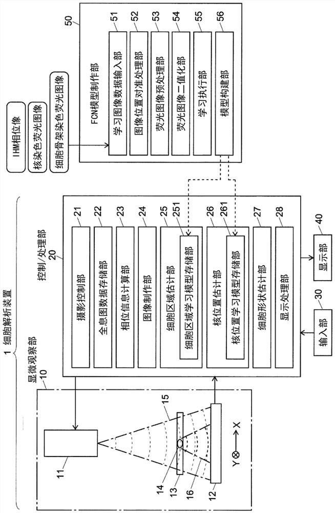
[0040] figure 1 It is a structural block diagram of main parts of a cell analysis device as an embodiment for carrying out the cell image analysis method according to the present invention.

[0041] The cell analysis device 1 of the present embodiment includes a microscopic observation unit 10 , a control / processing unit 20 , an input unit 30 as a user interface, and a display unit 40 . In addition, an FCN model creation unit 50 is attached to the cell analysis device 1 .

[0042] The microscopic observation unit 10 is an in-line holographic microscope (In-line Holographic Microscopy: IHM), and includes a light source unit 11 including a laser diode and the like and an image sensor 12, and a cell 14 is arranged between the light source unit 11 and the image sensor 12. Culture plate13.

[0...


PUM


Abstract

A cell image analysis method and a cell image analysis device are provided. A shape of a cell is favorably observed in a non-invasive manner. One aspect of the cell analysis device according to the present invention includes: holographic microscopy 10, an image generation unit 23,24 configured to generate a phase image of a cell based on hologram data acquired by observing the cell with the holographic microscopy, a first machine learning model storage unit 251 configured to store a cell area machine learning model generated by performing machine learning using training data in which a phase image of a cell generated based on the hologram data is used as an input image and a pseudo cell area image based on a stained image acquired by staining a cytoskeleton corresponding to the phase image is regarded as a ground truth image, and a cell area estimation unit 25 configured to output a cell area estimation image indicating a cell area using a machine learning model stored in the first machine learning model storage unit, wherein the phase image generated by the image generation unit for the analysis target cell is used as an input image.

Description

technical field [0001] The present invention relates to a cell image analysis method for analyzing and processing observed images of cells and a cell analysis device using the method, and more specifically, to a cell shape observation and cell area evaluation. A cell image analysis method and a cell analysis device. Background technique [0002] In the field of regenerative medicine, research using pluripotent stem cells such as iPS cells, ES cells, and mesenchymal stem cells has become popular in recent years. In research and development of regenerative medicine using such pluripotent stem cells, it is necessary to culture a large amount of undifferentiated cells maintaining pluripotency. Therefore, selection of an appropriate culture environment and stable control of the environment are required, and the state of cells in culture needs to be checked at high frequency. In the field of regenerative medicine, in order to continue culturing observed cells, it is important to...

Claims


Application Information

Patent Timeline
Patent Type & Authority Applications(China)
IPC IPC(8): G01N21/64C12Q1/02C12M1/34C12M1/00
CPCC12M41/36C12Q1/02G01N21/6402G01N21/6486G01N15/1429G01N15/1434G01N15/1463G01N15/1475G01N2015/1006G01N2015/1454G01N2015/1497G06T2207/10056G06T2207/20081G06T2207/20084G06T2207/30024G06T7/11G06T7/136G06T7/194G01N15/1468G01N2015/0065G01N2015/1477G06T7/0012G06T2207/30104
Inventor 赤泽礼子
Owner SHIMADZU SEISAKUSHO CO LTD
Why Eureka
Try Eureka