Magnetic resonance imaging method and system, and storage medium

A technology of magnetic resonance imaging and magnetic resonance imaging, which can be used in image enhancement, image analysis, measurement of magnetic variables, etc., and can solve the problems of low efficiency of magnetic resonance scanning.

Pending Publication Date: 2021-05-07
SHANGHAI UNITED IMAGING HEALTHCARE
View PDF9 Cites 2 Cited by
  • Summary
  • Abstract
  • Description
  • Claims
  • Application Information

AI Technical Summary

Problems solved by technology

[0004] Embodiments of the present invention provide a magnetic resonance imaging method, system, and storage

Method used


Image

  • Magnetic resonance imaging method and system, and storage medium
  • Magnetic resonance imaging method and system, and storage medium
  • Magnetic resonance imaging method and system, and storage medium

Examples


Embodiment 1

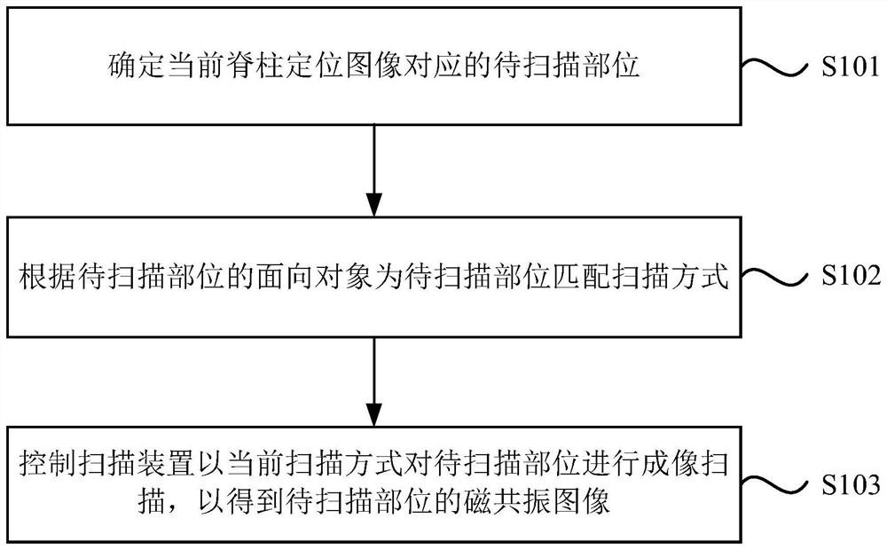
[0035] figure 1 It is a flow chart of the magnetic resonance imaging method provided by Embodiment 1 of the present invention. The technical solution of this embodiment is applicable to the situation of controlling the magnetic resonance system to automatically determine the part to be scanned and matching the scanning mode for the part to be scanned. The method can be executed by the magnetic resonance imaging device provided in the embodiment of the present invention, and the device can be implemented in the form of software and / or hardware, and configured to be applied in a processor. like figure 1 As shown, the method specifically includes the following steps:

[0036] S101. Determine the part to be scanned corresponding to the current spine positioning image.

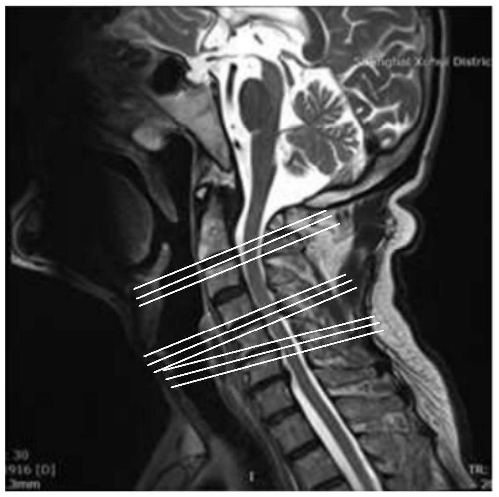
[0037] Among them, the spine includes vertebral bodies and intervertebral discs. Lesions in the spine are usually distributed over the vertebral bodies and / or intervertebral discs. During clinical magnetic res...

Embodiment 2

[0054] Figure 4 It is a flow chart of the magnetic resonance imaging method provided by Embodiment 2 of the present invention. On the basis of the foregoing embodiments, the embodiment of the present invention adds a step of switching the part to be scanned.

[0055] Correspondingly, the method of this embodiment includes:

[0056] S201. Determine the part to be scanned corresponding to the current spine positioning image.

[0057] S202. Match the scan mode for the part to be scanned according to the object orientation of the part to be scanned.

[0058] S203. Receive the user's request for switching the part to be scanned, complete the object-oriented switching of the part to be scanned according to the request for switching the part to be scanned, and match the corresponding scanning mode for the part to be scanned after switching.

[0059] If the user feels that the current part to be scanned is not suitable for the current patient, he will input a request to switch the...

Embodiment 3

[0066] Figure 5 It is a flow chart of the magnetic resonance imaging method provided by Embodiment 3 of the present invention. On the basis of any of the above-mentioned embodiments, the embodiment of the present invention adds a step of modifying the part to be scanned.

[0067] Correspondingly, the method of this embodiment includes:

[0068] S301. Determine the part to be scanned corresponding to the current spine positioning image.

[0069] S302. Match the scan mode for the part to be scanned according to the object orientation of the part to be scanned.

[0070] S303. Receive the user's request for modifying the range of the part to be scanned, and re-determine the range of the part to be scanned according to the range modification request to update the part to be scanned.

[0071] After the part to be scanned and its corresponding scanning mode are determined, if the user wants to modify the range of the part to be scanned currently, he inputs a range modification re...


PUM


Abstract

The embodiment of the invention discloses a magnetic resonance imaging method and system, and a storage medium. The method comprises the following steps of: determining a to-be-scanned position corresponding to a current spine positioning image; according to an oriented object of the to-be-scanned position, matching a scanning way for the to-be-scanned position; and controlling a scanning device to carry out imaging scanning on the to-be-scanned position according to the current scanning way to obtain a magnetic resonance image of the to-be-scanned position. By use of the method disclosed by the invention, the problem of low implementation efficiency of magnetic resonance scanning in the prior art is solved.

Description

technical field [0001] Embodiments of the present invention relate to the field of medical equipment, and in particular, to a magnetic resonance imaging method, system, and storage medium. Background technique [0002] When a lesion or injury occurs in the spine, it is usually necessary to perform an MR scan on the lesion or injury, so as to diagnose the lesion or injury of the spine through the MR image. However, before the scan starts, the doctor needs to determine the part to be scanned according to the spinal positioning image and the scanning method to scan the part to be scanned. This process is usually time-consuming and depends on the doctor's experience. [0003] Therefore, it is necessary to provide a magnetic resonance imaging method to improve the implementation efficiency of the magnetic resonance imaging scan. Contents of the invention [0004] Embodiments of the present invention provide a magnetic resonance imaging method, system, and storage medium to sol...

Claims


Application Information

Patent Timeline
IPC IPC(8): A61B5/055
CPCA61B5/055G01R33/4835G01R33/543G01R33/5602A61B5/0035A61B5/4566A61B5/7264A61B6/037A61B6/505A61B6/5247A61B6/545G06T7/0012G01R33/5608G01R33/546G06T2200/24G06T2207/30012
Inventor 辛阳史庭荣
Owner SHANGHAI UNITED IMAGING HEALTHCARE
Why Eureka
Try Eureka