
A Quantitative Evaluation System for Ultrasonic Images

An ultrasound image, quantitative evaluation technology, applied in ultrasound/sonic/infrasound image/data processing, image enhancement, image analysis, etc. Problems such as the depth of cartilage

Active Publication Date: 2022-05-24
TSINGHUA UNIV
View PDF11 Cites 0 Cited by
  • Summary
  • Abstract
  • Description
  • Claims
  • Application Information

AI Technical Summary

Problems solved by technology

However, for clinical ultrasonography, due to the compromise between probe frequency and imaging depth, high-frequency probes used in ex vivo experiments or arthroscopy cannot detect the depth of articular cartilage, so they are not suitable for clinical examination of articular cartilage
Moreover, most of the ultrasound machines currently used in clinical practice cannot provide ultrasound radio frequency data
Therefore, the indicators commonly used in ex vivo experiments and arthroscopy to reflect the microstructural changes of the cartilage surface and the indicators extracted from the frequency spectrum analysis of ultrasound radio frequency data cannot be applied to clinical ultrasound examination.

Method used


Image

  • A Quantitative Evaluation System for Ultrasonic Images
  • A Quantitative Evaluation System for Ultrasonic Images
  • A Quantitative Evaluation System for Ultrasonic Images

Examples


Embodiment Construction

[0046] In order to make the objectives, technical solutions and advantages of the present invention clearer, the technical solutions of the present invention will be clearly and completely described below with reference to the accompanying drawings. Obviously, the described embodiments are some, but not all, embodiments of the present invention. Based on the embodiments of the present invention, all other embodiments obtained by those of ordinary skill in the art without creative efforts shall fall within the protection scope of the present invention.

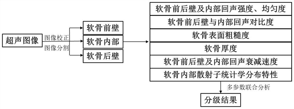
[0047] like figure 1 As shown, the ultrasonic image quantitative evaluation method provided by this embodiment includes the following steps:

[0048] 1. The steps of preprocessing the acquired ultrasound images:

[0049] In this embodiment, the acquired ultrasonic image is a joint ultrasonic image in a standard format (such as DICOM format) acquired by an ultrasonic system according to a unified standard operating procedure (...


PUM


Abstract

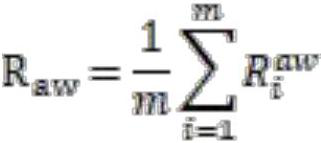
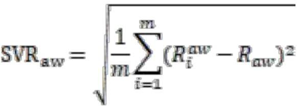
The present invention relates to an ultrasound image quantitative evaluation system, comprising: an image preprocessing module for preprocessing acquired joint ultrasound images; an image segmentation module for image segmentation of joint ultrasound images; a quantitative analysis index extraction module, It is used for quantitative analysis index extraction of joint ultrasound images; the parameter joint analysis module is used for multi-parameter joint analysis of joint ultrasound images. The present invention automatically segments the front and rear walls and internal regions of cartilage on the ultrasonic image, and conducts quantitative analysis on the segmented regions of interest, extracts information related to ultrasonic echo intensity, sound attenuation, cartilage thickness, and scattering sub-statistical distribution. Quantitative analysis indicators, and then use the method of multivariate analysis to comprehensively analyze various indicators, so as to realize the non-invasive and quantitative evaluation of the degree of articular cartilage lesions.

Description

technical field [0001] The invention relates to an ultrasonic image quantitative evaluation system, in particular to an ultrasonic image quantitative evaluation system that can be used to evaluate articular cartilage lesions. Background technique [0002] Osteoarthritis is the most common chronic degenerative joint disease in the elderly. Early identification of osteoarthritis and accurate grading of joint degeneration facilitate timely and appropriate interventions to control disease progression and significantly improve prognosis. Clinically, conventional radiography (CR) is often used to evaluate bone and joint structural changes, but it cannot show soft tissue lesions. Magnetic resonance imaging (MRI) can be used to detect articular cartilage, synovium, meniscus, and soft tissue lesions, but it is expensive, takes a long time to scan, and is insensitive to early cartilage lesions. Ultrasound diagnosis has the advantages of real-time, portable, easy-to-use, and low cost...

Claims


Application Information

Patent Timeline
Patent Type & Authority Patents(China)
IPC IPC(8): A61B8/08A61B8/00G06T7/11
CPCA61B8/0875A61B8/5215G06T7/11G06T2207/10132G06T2207/30008
Inventor 罗建文王媛媛何琼高孟泽
Owner TSINGHUA UNIV
Learn More