Assays for cell-based therapies or treatments
A cellular, therapeutic technique, applied in the field of assays for cell-based therapy or therapy, that addresses difficulties, time consuming, lack of quantitative results, etc.
- Summary
- Abstract
- Description
- Claims
- Application Information
AI Technical Summary
Problems solved by technology
Method used
Image
Examples
Embodiment 1
[0098] Example 1 - Coated and Uncoated Substrates
[0099] Different coating agents were tested to determine suitable substrates for the methods of the present disclosure.
[0100] Opaque 96-well assay plates were uncoated or coated with poly-D-lysine or coated with human fibronectin. Plates were coated with poly-D-lysine by incubating 25 μl to 50 μl of a 200 μg / ml poly-D-lysine solution in each well for five minutes to two hours at room temperature. Alternatively, 250 μl of a 200 μg / ml poly-D-lysine solution was incubated in each well for one hour at room temperature. After incubation, the plates were washed with ddH 2 O rinse and then left to air dry for two hours. Plates were coated with human fibronectin by incubating 300 μl of a 20 μg / ml human fibronectin solution overnight at 37°C. After overnight incubation, plates were rinsed with Advanced DMEM / F12.
[0101] Human retinoblastoma (hRB) Y79 ( HTB-18 TM ) cells were added to three different assay plates. In the c...
Embodiment 2
[0102] Example 2 - Fluorescence-based cell viability assay
[0103] Fluorescence-based assays that measure cell viability were tested for use in the methods of the present disclosure.
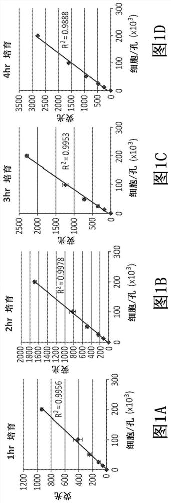
[0104] First, the 12.5×10 3 , 2.5×10 4 , 5.0×10 4 , 1.0×10 5 and 2.0×10 5 Individual human retinoblastoma (hRB) Y79( HTB-18 TM ) cells were seeded in five separate wells of a 96-well assay plate and incubated in 50 μl of SM. After allowing the cells to settle for 30 minutes to two hours, the cells were then incubated after being thoroughly mixed with 250 μl of human retinoblastocyte conditioned medium. Then 200 μl of medium was withdrawn and 20 μl of diluted resazurin (7-hydroxy-3H-phenoxazin-3-one 10-oxide sodium salt) was added to the cells at 37°C Reagents (1:4 in DPBS) lasted 1, 2, 3 or 4 hours. Resazurin is a compound that reduces to a compound called resorufin when internalized by living cells. Resorufin is highly fluorescent, meaning that the fluorescent signal produced can b...
Embodiment 3
[0106] Example 3 - Determining the potency of human retinoblastoid conditioned medium
[0107] The methods of the present disclosure were used to test the potency of human retinoblastocyte conditioned medium.
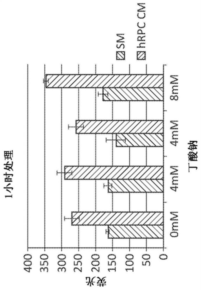
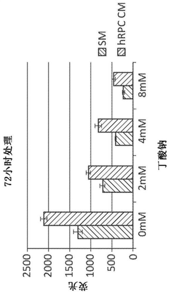
[0108] First, take a 2.5×10 4 hRB cells were seeded on the assay plate at a density of 2 cells / well. Cells were then incubated with human retinoblastocyte conditioned medium (hRPC CM) or standard medium (SM; standard human retinoblastoid medium that has not been exposed to human retinal progenitor cells). Media were not supplemented with sodium butyrate, supplemented with 2 mM sodium butyrate, 4 mM sodium butyrate or 8 mM sodium butyrate. Freshly made 200 mM sodium butyrate was added to 250 μl of medium to generate various sodium butyrate concentrations. Use 9.0×10 6 Individual human retinal progenitor cells generate hRPC CMs.
[0109] hRB cells were incubated in 300 μl of each medium for one hour or 72 hours. After incubation, use as described above Reagents to...
PUM
 Abstract
Description
Claims
Application Information
 - R&D
- Intellectual Property
- Life Sciences
- Materials
- Tech Scout
- Unparalleled Data Quality
- Higher Quality Content
- 60% Fewer Hallucinations
Browse by: Latest US Patents, China's latest patents, Technical Efficacy Thesaurus, Application Domain, Technology Topic, Popular Technical Reports.
© 2025 PatSnap. All rights reserved.Legal|Privacy policy|Modern Slavery Act Transparency Statement|Sitemap|About US| Contact US: help@patsnap.com


